Comparative Study
. 2008 Jan 18;375(3):637-49.
doi: 10.1016/j.jmb.2007.10.006. Epub 2007 Oct 10.

Prevention of cardiac hypertrophy and heart failure by silencing of NF-kappaB

Affiliations
Comparative Study

Prevention of cardiac hypertrophy and heart failure by silencing of NF-kappaB

Sudhiranjan Gupta et al. J Mol Biol. .

Abstract

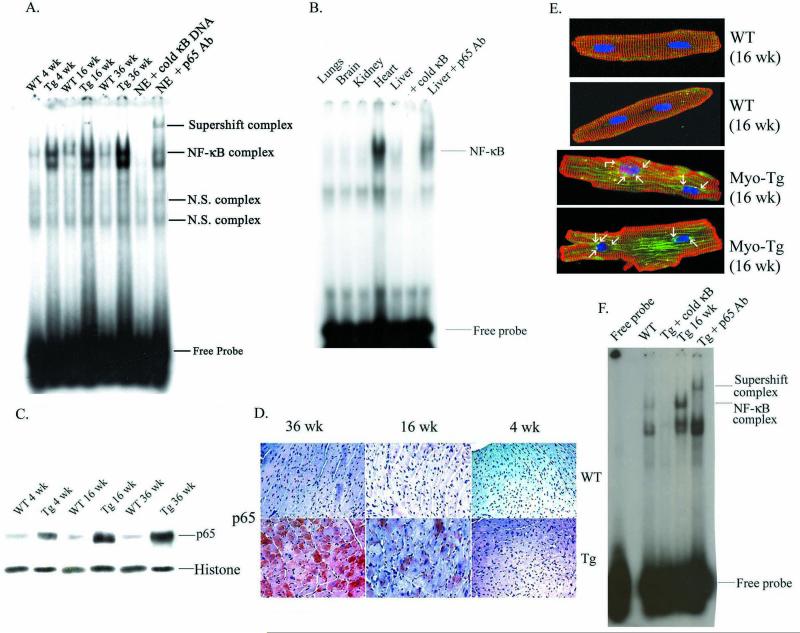
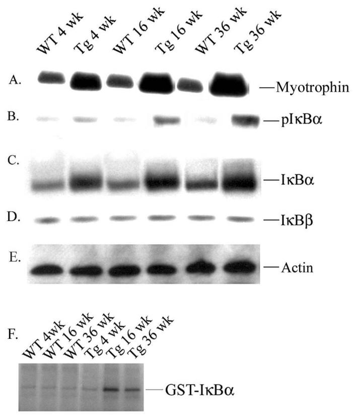
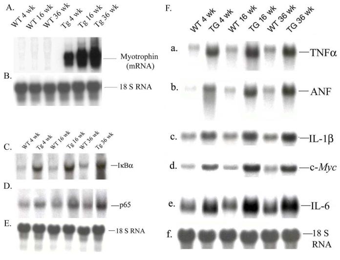
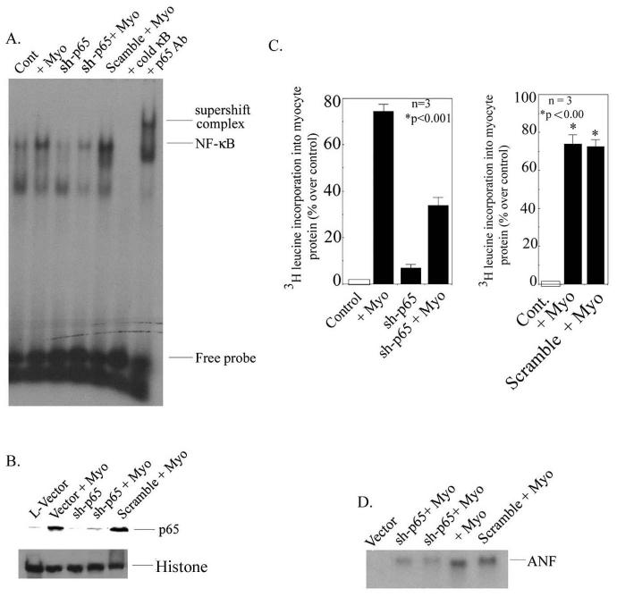
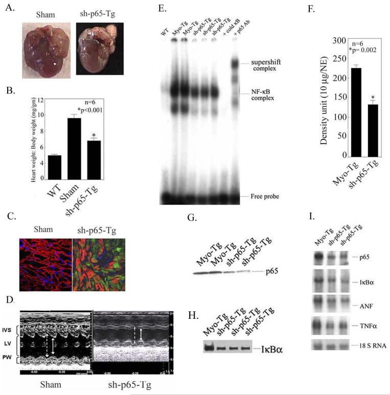
Activation of the nuclear factor (NF)-kappaB signaling pathway may be associated with the development of cardiac hypertrophy and its transition to heart failure (HF). The transgenic Myo-Tg mouse develops hypertrophy and HF as a result of overexpression of myotrophin in the heart associated with an elevated level of NF-kappaB activity. Using this mouse model and an NF-kappaB-targeted gene array, we first determined the components of NF-kappaB signaling cascade and the NF-kappaB-linked genes that are expressed during the progression to cardiac hypertrophy and HF. Second, we explored the effects of inhibition of NF-kappaB signaling events by using a gene knockdown approach: RNA interference through delivery of a short hairpin RNA against NF-kappaB p65 using a lentiviral vector (L-sh-p65). When the short hairpin RNA was delivered directly into the hearts of 10-week-old Myo-Tg mice, there was a significant regression of cardiac hypertrophy, associated with a significant reduction in NF-kappaB activation and atrial natriuretic factor expression. Our data suggest, for the first time, that inhibition of NF-kappaB using direct gene delivery of sh-p65 RNA results in regression of cardiac hypertrophy. These data validate NF-kappaB as a therapeutic target to prevent hypertrophy/HF.


Figures

Figure 1
Figure 1
NF-κB activation during progression of hypertrophy in Myo-Tg mice hearts. (A) Nuclear protein was extracted from the hearts of 4-, 16-, and 36-week-old WT and Myo-Tg mice. Binding reactions were performed with an NF-κB oligonucleotide labeled with 32P-dATP. The complex formation was eliminated with excess unlabeled NF-κB oligonucleotide. The complex formation was confirmed by supershift analysis using p65 antibody. NE: Nuclear extract from 16 week Myo-Tg mice. (B) Nuclear protein was extracted from lung, brain, kidney, heart and liver separately and binding reactions were performed as described in (A). (C) Western blots profile of NF-κB p65 protein into the nucleus. Histone antibody was used as an internal nuclear protein loading control. (D) Expression of p65 active protein in the heart section of both WT and Myo-Tg mice and were photographed with an Olympus photomicroscope at 20 X magnification. This figure is representative of six different mice in each group (WT and Myo-Tg). (E). Confocal images of p65 protein from adult myocytes taken from WT and Myo-Tg mice at 16 weeks of age and were stained with p65 polyclonal antibody followed by FITC-conjugate (green), alpha-sarcomeric actinin monoclonal antibody for myocyte staining (red), followed by Texas Red-conjugate and DAPI (blue) for nuclear staining. Immunofluorescent NF-κB p65 staining was observed using a laser scanning confocal microscope and 63 X, 0.7 oil immersion objective lens. The figure is a representative of 3 different mice. The arrow indicates the translocation of NF-κB-p65 orotein into the nucleus. (F) EMSA analysis using nuclear extract from adult myocytes from 16-week old Myo-Tg mice.
Figure 2
Figure 2
Expression of myotrophin, IκBα proteins and IKKβ activity in the hearts of Myo-Tg mice. Cytoplasmic protein extracts were made from both WT and Myo-Tg mouse hearts at 4, 16, and 36 weeks of age. Tissue extracts (10 μg) were analyzed for (A) myotrophin and tissue extracts (50 μg) were analyzed for (B) IκBα phosphorylation, (C) the intracellular level of total IκBα protein content, and (D) IκBβ level using myotrophin, phospho-IκBα, IκBα, and IκBβ antibody as probes. (E) Actin protein was used as an internal loading control. Results are presented as the mean SEM and represent six different mice in each group (WT and Myo-Tg) (p < 0.001 compared with the WT mice). (F) Tissue extracts (500 μg) were immuno-precipitated with IKKβ antibody and kinase activity was determined with GST-IκBα as a substrate. These results are presented as the mean SEM and represent six different mice (p < 0.001 compared with the WT mice).
Figure 3
Figure 3
Transcriptional analysis of myotrophin, IκBα, p65, and NF-κB-dependent genes during progression of cardiac hypertrophy. Total RNA was extracted from hearts of 4-, 16-, and 36-week old WT and Myo-Tg mice. mRNA expression was determined using (A) Myotrophin (C) IκBα and (D) p65 cDNAs labeled with 32P-dCTP as a probes. (B and E) 18S rRNA probe was used as a loading control. Results are presented as the mean SEM and represent five different mice (p < 0.001 compared with the WT mice). (F). NF-κB-dependent gene expression was performed by northern blot using (a) TNFα, (b) ANF, (c) IL-1β, (d) IL-6, (e) c-myc cDNAs as a probes; 18S rRNA was used as an internal loading control. These results are presented as the mean SEM and represent three different mice (p < 0.001 compared with the WT mice).
Figure 4
Figure 4
RNA interference suppresses NF-κB-p65 activation, myocyte growth, and ANF expression in neonatal myocytes. (A) Cells were transduced with L-sh-p65 at a multiplicity of infection (MOI) of 30 for 48 hours and were stimulated with myotrophin. EMSA was performed as described in Fig 1. (B) Western blot analysis using NF-κB-p65 antibody as a probe in L-sh-p65-transduced neonatal myocytes. L-EGFP was used as a control. Histone antibody was used as an internal nuclear protein loading control. (C). Myocyte growth was measured by [3H]-leucine incorporation into myocyte protein. L-sh-p65 was transduced into neonatal myocytes in presence and absence of myotrophin. Cells were also transduced with scramble sequences separately in presence and absence of myotrophin. Results are presented as the mean ± SEM and represent set of three different experiments in neonatal myocytes (p < 0.001 compared with unstimulated cells). (D) ANF expression was measured by using ANF cDNA as a probe in L-sh-p65-transduced neonatal myocytes in the presence or absence of myotrophin. Note: in all experiments, a scrambled sequence did not show any effect.
Figure 5
Figure 5
Attenuation of cardiac mass and inhibition of NF-κB activation cascade in Myo-Tg mice treated with L-sh-p65. (A) Typical appearance of heart size in untreated Myo-Tg and L-sh-p65-transduced Myo-Tg mice. (B) Heart weight:body weight ratio in L-sh-p65-transduced Myo-Tg mice. Values represent mean ± SE and p = 0.002 compared with untreated Myo-Tg mice (n=6). (C) Confocal image showing the expression of EGFP in L-sh-p65-transduced Myo-Tg mice. Magnification is 40 X. (D) Representative M-mode tracings of the left ventricle obtained in a Myo-Tg (Sham) and sh-p65-Tg-treated mice. Arrows indicate endocardial borders in diastole (broken arrows) and systole (full arrows). Improved LV fractional shortening and decreased left ventricle internal dimension are evident in the L-sh-p65-treated mouse. IVS, interventricular septum; LV, left ventricle cavity; PW, posterior wall. (E) EMSA was performed using nuclear extract from WT, Myo-Tg, and L-sh-p65-treated Myo-Tg mice as described in Fig. 1A. (E) Quantification of EMSA using an arbitrary density unit (10 μg/NE). NE: nuclear extract. These results are presented as the mean ± SE and represent six different mice (p = 0.002 compared with the sham mice, two-way ANOVA). (G) p65 nuclear protein translocation was demonstrated by western blot analysis p65 antibody as described in Fig. 1B. (H) IκBα protein levels were determined by western blot analysis using IκBα antibody. (I) mRNA expression profiling of IκBα, p65, ANF, and TNFα in Myo-Tg and L-sh-p65-transduced Myo-Tg mice (two separate animals) using specific cDNAs as probes.

References

    1. Levy D, Garrison RJ, Savage DD, Kannel WB, Castelli WP. Prognostic implications of echocardiographically determined left ventricular mass in the Framingham Heart Study. N Engl J Med. 1990;322:1561–1566. - PubMed
    1. Kent RL, Mann DL, Cooper G. Signals for cardiac muscle hypertrophy in hypertension. J Cardiovasc. Pharmacol. 1991;17(Suppl 2):S7–13. - PubMed
    1. Baldwin AS., Jr. The NF-κB and I-κB proteins: new discoveries and insights. Annu Rev Immunol. 1996;14:649–683. - PubMed
    1. May MJ, Ghosh S. Signal transduction through NF-kappa B. Immunol Today. 1998;19:80–88. - PubMed
    1. Chen F, Castranova V, Shi X, Demers LMA. New insights into the role of nuclear factor-κB, a ubiquitous transcription factor in the initiation of diseases. Clin. Chem. 1999;45:7–17. - PubMed

Publication types

MeSH terms