. 2008 Jan 18;375(3):720-34.
doi: 10.1016/j.jmb.2007.10.064. Epub 2007 Nov 1.

Restriction endonuclease inhibitor IPI* of bacteriophage T4: a novel structure for a dedicated target

Affiliations

Restriction endonuclease inhibitor IPI* of bacteriophage T4: a novel structure for a dedicated target

Dalin Rifat et al. J Mol Biol. .

Abstract

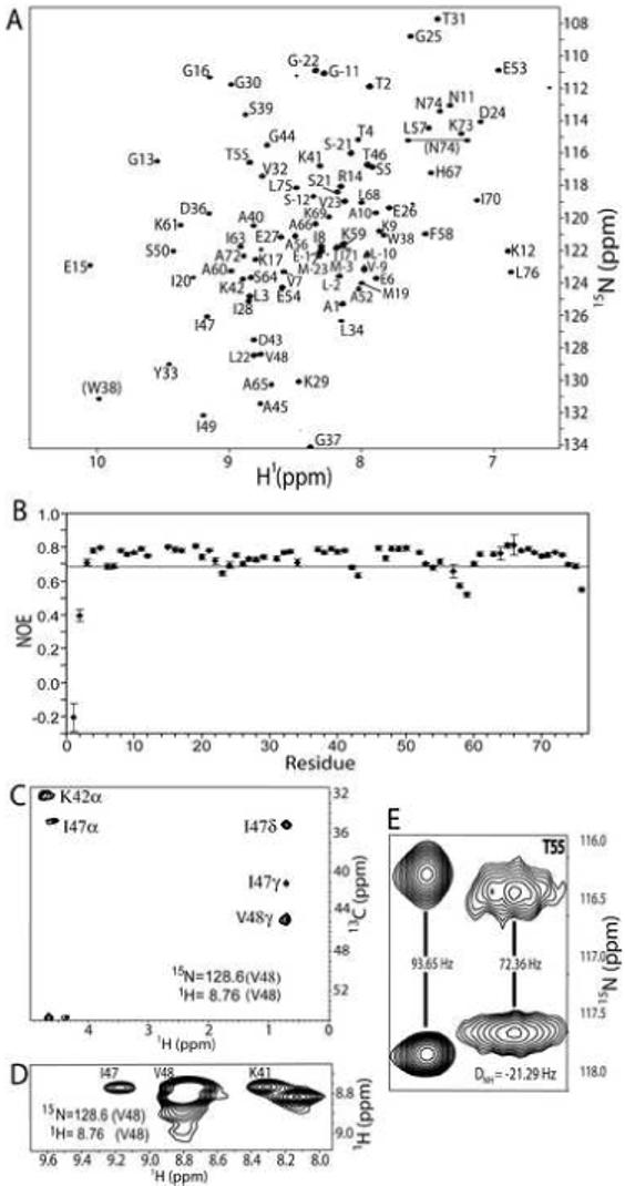
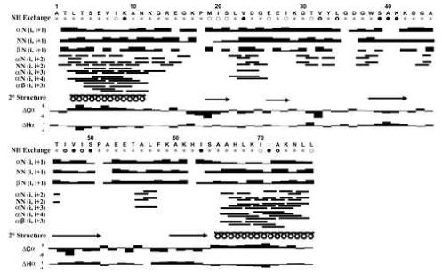
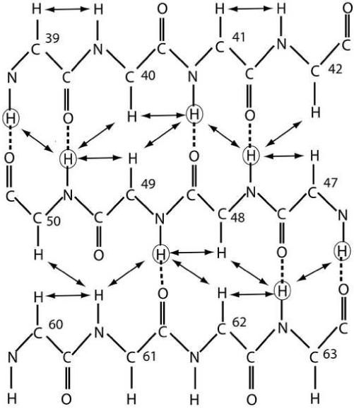
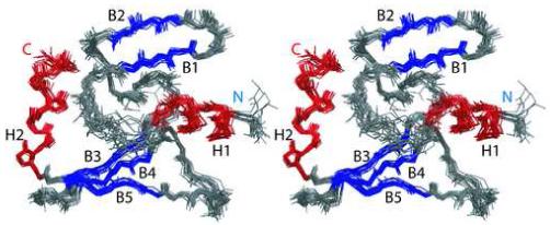
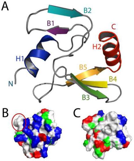
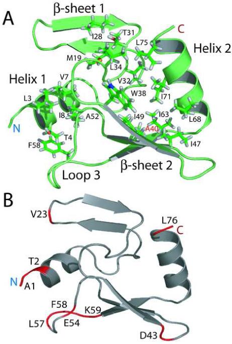
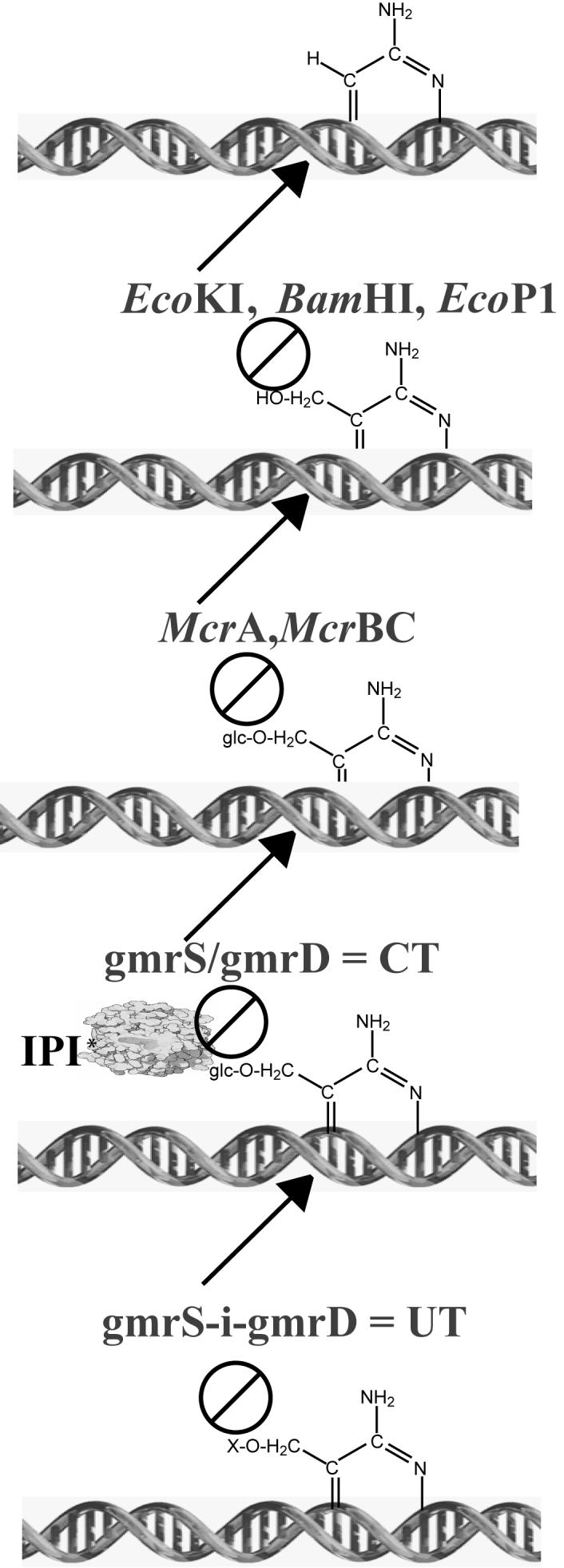
Phage T4 protects its DNA from the two-gene-encoded gmrS/gmrD (glucose-modified hydroxymethylcytosine restriction endonuclease) CT of pathogenic Escherichia coli, CT596, by injecting several hundred copies of the 76-amino-acid-residue nuclease inhibitor, IPI*, into the infected host. Here, the three-dimensional solution structure of mature IPI* is reported as determined by nuclear magnetic resonance techniques using 1290 experimental nuclear Overhauser effect and dipolar coupling constraints ( approximately 17 constraints per residue). Close examination of this oblate-shaped protein structure reveals a novel fold consisting of two small beta-sheets (beta1: B1 and B2; beta2: B3-B5) flanked at the N- and C-termini by alpha-helices (H1 and H2). Such a fold is very compact in shape and allows ejection of IPI* through the narrow 30-A portal and tail tube apertures of the virion without unfolding. Structural and dynamic measurements identify an exposed hydrophobic knob that is a putative gmrS/gmrD-binding site. A single gene from the uropathogenic E. coli UT189, which codes for a gmrS/gmrD-like UT fusion enzyme (with approximately 90% identity to the heterodimeric CT enzyme), has evolved IPI* inhibitor immunity. Analysis of the gmrS/gmrD restriction endonuclease enzyme family and its IPI* family phage antagonists reveals an evolutionary pathway that has elaborated a surprisingly diverse and specifically fitted set of coevolving attack and defense structures.


Figures

Figure 1
Figure 1
(A) 2D 1H-15N HSQC of IPI* at 600 MHz (1H) with residue assignments included. Backbone 1H-15N correlations are labeled sequence-specifically, and correlations connected by the horizontal line correspond to asparagine side-chain NH2 groups. (B) A graph representing data obtained from a 2D 15N-{1H} heteronuclear NOESY experiment shows that A1, T2, V23, D43, L57, F58, K59, and L76 all have significant fast-motion contributions (NOE<0.68; residues below the horizontal line in graph). A plane from the 4D 13C, 15N-edited NOESY-HSQC (C) and a 3D 15N-edited NOESY (D) show NOE correlations of the amide hydrogen of V48 to sequential and long-range resonances. In particular, NOE correlations of HαHN (i, i+1), HαHN (K42, V48), HNHN (K41, V48) are visible. The presence of a strong HαHN (i, i+1) resonance and strong long-range resonances is indicative of a residue in the middle of a beta sheet. (E) A representative region of the 2D 1H-15N HSQC IPAP NMR spectra used to measure dipolar coupling data (RDCs). This region illustrates splitting of T55. Dipolar couplings were determined by comparing the splitting in the presence (right) or absence (left) of the aligning acrylamide media.
Figure 2
Figure 2
Secondary structure of IP1. Circles indicate relative amide hydrogen exchange rates at 37°C as determined using 2D 1H-15N HSQC spectra with stars (T<0.125h) and open circles (0.125h <T<0.5 h) arbitrarily referred to as fast exchanging amide protons. Residues with partially filled in circles are arbitrarily referred to as medium exchanging amide protons (0.5<T<1 h). Residues with solid circles (T>4 h) are arbitrarily referred to as slowly exchanging amide protons. The NOE correlations were determined from 3D 15N-edited NOESY-HSQC, 4D 13C, 15N-edited NOESY, and 4D 13C, 13C-edited NOESY at 37°C. The height of the bar indicated the strength of the NOE (strong, medium, medium-weak, weak, or very weak). Deviations in the 13Cα and 1Hα chemical shift from those of a random coil are illustrated such that regions of contiguous upfield-shifted 13Cα chemical shifts (positive values) and downfield-shifted 1Hα chemical shifts (negative values) are indicative of helical regions. Likewise, regions of contiguous downfield-shifted 13Cα chemical shifts (negative values) and upfield-shifted 1Hα chemical shifts (positive values) are indicative of regions of β-strand. The secondary structure is represented by α-helices (spirals), β-strands (arrows), turns (crosses), loops and flexible regions (no symbols), as indicated under the appropriate residues in the sequence of IPI*.
Figure 3
Figure 3
Schematic of the 3-strand antiparallel beta sheet. Unambiguous NOEs are marked by solid arrows. Circled hydrogen atoms are those involved in slow exchange with the solution, and dotted lines represent hydrogen bonds.
Figure 4
Figure 4
Stereoview of the 20 lowest energy structures of IPI*. Helices are colored red, beta strands are colored blue, turns and loops and flexible regions are colored gray.
Figure 5
Figure 5
NMR solution structure of IPI*. (A) Ribbon diagram of the lowest-energy structure of IPI*. (B) and (C) The electrostatic surface potential of IP1. Electronegative (acidic) regions are colored red, electropositive (basic) regions are colored blue, uncharged regions are colored white and polar group residues are colored green. In (B), exposed hydrophobic patch residues 55-58 are circled in red.
Figure 6
Figure 6
Hydrophobic residues involved in the unique fold of IPI*. (A) IPI* maintains its fold through interactions within a large hydrophobic fold. Residues included in this hydrophobic pocket are L3, T4, V7, and I8 on helix 1, M19 and I28 on the first β-sheet, T31, V32, and L34 in loop 2, W38, A40, I47, I49, and I63 in the second β-sheet, A52 and F58 in the loop 3, and L68, I71, and L75 in helix 2. Labeled in red is residue A40, where an A40T substitution eliminates IPI* structure and inhibitor function. (B) Ribbon diagram illustrating residues (in red) that exhibit fast time-scale motions. Several of the residues with significant fast-motion contributions are located in loop 3.
Figure 7
Figure 7
Evolution of the T-even phage IPI* inhibitors and DNA modifications in response to type IV DNA modification dependent restriction endonucleases. Bacterial encoded type I, II, and III (e.g. E. coli K12, BamHI, P1) restriction endonucleases protect against infecting phage DNAs containing cytosine (first line). Many of these enzymes are blocked by 5-methyl or 5-hydroxymethylcytosine (HMC) DNA modification (line 2). The McrA and McrBC type IV modification dependent restriction endonucleases of E. coli specifically attack HMC modified DNAs, but are inhibited by the glucosylation of HMC (glc-HMC) (line 3). The heterodimeric CT (gmr (glucose modified restriction) enzyme of E. coli CT596 hydrolyzes only the sugar modified (glc)-HMC containing DNAs of a number of T-even phages, but its activity is inhibited by the encapsidated phage IPI* protein injected with the DNA (line 4). The UT enzyme comprising the S and D subunits in a single polypeptide chain is immune to IPI*, but is blocked by further unknown modifications to the DNAs or internal proteins of some of the T-even phages (line 5).

References

    1. Abremski K, Black LW. The function of bacteriophage T4 internal protein I in a restrictive strain of Escherichia coli. Virology. 1979;97(2):439–47. - PubMed
    1. Bair CL, Rifat D, Black LW. Exclusion of glucosyl-hydroxymethylcytosine DNA containing bacteriophages is overcome by the injected protein inhibitor IPI*. J Mol Biol. 2007;366(3):779–89. - PMC - PubMed
    1. Bair CL, Black LW. A type IV modification dependent restriction nuclease that targets glucosylated hydroxymethyl cytosine modified DNAs. J Mol Biol. 2007;366(3):768–78. - PMC - PubMed
    1. Repoila F, Tetart F, Bouet JY, Krisch HM. Genomic polymorphism in the T-even bacteriophages. The EMBO journal. 1994;13(17):4181–92. - PMC - PubMed
    1. Mullaney JM, Black LW. Capsid targeting sequence targets foreign proteins into bacteriophage T4 and permits proteolytic processing. J Mol Biol. 1996;261(3):372–85. - PubMed

Publication types

MeSH terms

Associated data