. 2007 Dec;9(12):1419-27.
doi: 10.1038/ncb1661. Epub 2007 Nov 25.

MUC1 oncoprotein activates the IkappaB kinase beta complex and constitutive NF-kappaB signalling

Affiliations

MUC1 oncoprotein activates the IkappaB kinase beta complex and constitutive NF-kappaB signalling

Rehan Ahmad et al. Nat Cell Biol. 2007 Dec.

Abstract

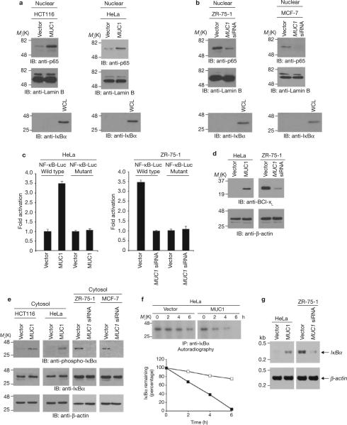
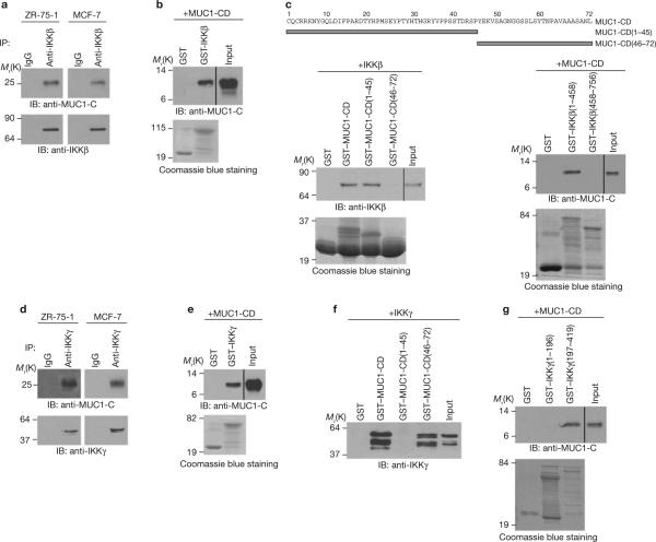
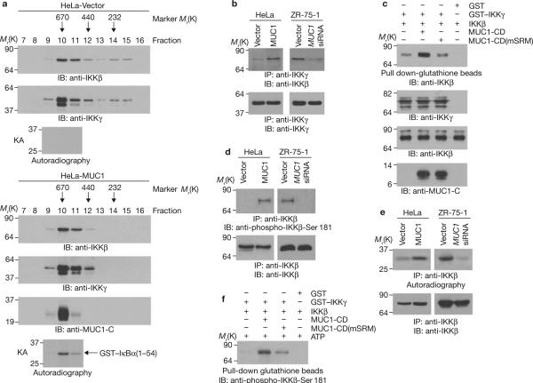
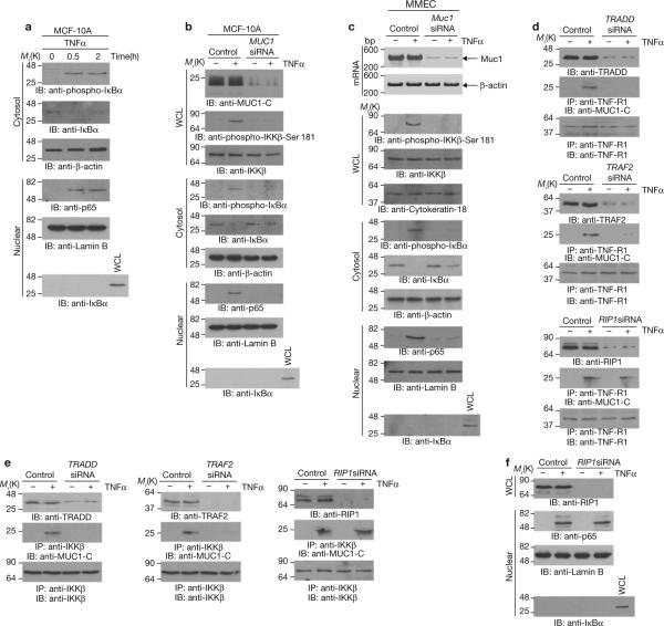
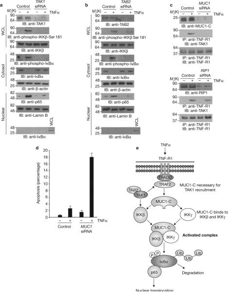
Nuclear factor-kappaB (NF-kappaB) is constitutively activated in diverse human malignancies by mechanisms that are not understood. The MUC1 oncoprotein is aberrantly overexpressed by most human carcinomas and, similarly to NF-kappaB, blocks apoptosis and induces transformation. This study demonstrates that overexpression of MUC1 in human carcinoma cells is associated with constitutive activation of NF-kappaB p65. We show that MUC1 interacts with the high-molecular-weight IkappaB kinase (IKK) complex in vivo and that the MUC1 cytoplasmic domain binds directly to IKKbeta and IKKgamma. Interaction of MUC1 with both IKKbeta and IKKgamma is necessary for IKKbeta activation, resulting in phosphorylation and degradation of IkappaBalpha. Studies in non-malignant epithelial cells show that MUC1 is recruited to the TNF-R1 complex and interacts with IKKbeta-IKKgamma in response to TNFalpha stimulation. TNFalpha-induced recruitment of MUC1 is dependent on TRADD and TRAF2, but not the death-domain kinase RIP1. In addition, MUC1-mediated activation of IKKbeta is dependent on TAK1 and TAB2. These findings indicate that MUC1 is important for physiological activation of IKKbeta and that overexpression of MUC1, as found in human cancers, confers sustained induction of the IKKbeta-NF-kappaB p65 pathway.


Figures

Figure 1
Figure 1
MUC1 targets NF-κB p65 to the nucleus by inducing phosphorylation and degradation of IκBα. (a) and (b) Nuclear lysates from the indicated cells were subjected to immunoblotting with anti-p65, anti-lamin B and anti-IκBα antibodies. Whole cell lysate (WCL) prepared from HCT116-vector cells was used as a control for anti-IκBα reactivity. Immunoblot analysis of the nuclear lysates with antibodies against nuclear lamin B and cytosolic IκBα confirmed equal loading of the lanes and lack of cytoplasmic contamination. (c) The indicated cells were transfected with a pNF-κB-Luc reporter plasmid or a mutant at the NF-κB binding site and, as a control, the SV40-Renilla-Luc plasmid. Luciferase activity was measured at 48 h after transfection. The results are expressed as the fold activation (mean ± s.d., of three separate experiments) compared with that obtained in HeLa-vector (left) or ZR-75-1-MUC1 siRNA (right) cells (each assigned a value of 1). (d) Whole cell lysates from the indicated cells were immunoblotted with anti-Bcl-xL and anti-β-actin antibodies. (e) Cytosolic lysates from the indicated cells were immunoblotted with anti-phospho-IκBα, anti-IκBα and anti-β-actin antibodies. (f) HeLa-vector and HeLa-MUC1 cells were pulsed with 35S-methionine and chased for the indicated times. Anti-IκBα immunoprecipitates from equal amounts of lysate were subjected to SDS–PAGE and autoradiography (upper panels). Intensity of the IκBα signals was determined by scanning densitometry and is expressed as the percentage IκBα remaining compared with that obtained at 0 h (lower panels). Similar results were obtained in two separate experiments. (g) IκBα and β-actin mRNA levels were determined for the indicated cells by quantitative RT-PCR. Full scans of the gels in a, b, e and f are shown in Supplementary Fig. S6-1.
Figure 2
Figure 2
MUC1-CD binds directly to IKKβ and IKKγ. (a) Lysates from the indicated cells were subjected to immunoprecipitation with a control IgG or anti-IKKβ antibody. The precipitates were immunoblotted with the indicated antibodies. (b) GST and GST–IKKβ bound to glutathione–agarose beads were incubated with purified MUC1-CD. The precipitates and input were immunoblotted with an anti-MUC1-C antibody. Input of the GST and GST–IKKβ proteins was assessed by Coomassie blue staining. (c) Amino-acid sequence of MUC1-CD (upper panel). GST and the indicated GST–MUC1-CD fusion proteins bound to glutathione beads were incubated with purified IKKβ. The precipitates and input were immunoblotted with an anti-IKKβ antibody (lower left). GST and the indicated GST–IKKβ fusion proteins bound to glutathione beads were incubated with MUC1-CD. The precipitates and input were immunoblotted with an anti-MUC1-C antibody (lower right). Input of GST and GST– fusion proteins was assessed by Coomassie blue staining. (d) Lysates from the indicated cells were subjected to immunoprecipitation with a control IgG or an anti-IKKγ antibody. The precipitates were immunoblotted with the indicated antibodies. (e) GST and GST–IKKγ bound to glutathione beads were incubated with purified MUC1-CD. The precipitates and input were immunoblotted with an anti-MUC1-C antibody. (f) GST and the indicated GST–MUC1-CD fusion proteins bound to glutathione beads were incubated with purified IKKγ. The precipitates and input were immunoblotted with an anti-IKKγ antibody. Input of the GST and GST– fusion proteins is shown in Fig. 2c, left. (g) GST and the indicated GST–IKKγ fusion proteins bound to glutathione beads were incubated with MUC1-CD. The precipitates and input were immunoblotted with an anti-MUC1-C antibody. Full scans of the gels in a, b, c, e and f and g are shown in Supplementary Fig. S6-2.
Figure 3
Figure 3
MUC1 activates the IKKβ–IKKγ complex. (a) HeLa-vector and HeLa-MUC1 cell lysates were separated on a Sephacryl S-200 HR column. The indicated fractions were analysed by immunoblotting with the indicated antibodies and for phosphorylation of GST–IκBα in kinase assays (KAs). (b) Anti-IKKγ immunoprecipitates from the indicated cells were immunoblotted with anti-IKKβ and anti-IKKγ antibodies. (c) GST or GST–IKKγ bound to glutathione beads was incubated with IKKβ in the absence and presence of MUC1-CD or MUC1-CD(mSRM). The precipitates were immunoblotted with an anti-IKKβ antibody (upper panel). Input of the proteins was assessed by immunoblotting with the indicated antibodies (lower 3 panels). (d) Anti-IKKβ precipitates from the indicated cells were immunoblotted with anti-phospho- IKKβ-Ser 181 and anti-IKKβ antibodies. (e) Anti-IKKβ precipitates from the indicated cells were incubated with GST-IκBα(1–54) and γ-32P-ATP. The reaction products were analysed by SDS–PAGE and autoradiography (upper panels). The precipitates were also immunoblotted with an anti-IKKβ antibody (lower panels). (f) GST or GST–IKKγ bound to glutathione beads was incubated with IKKβ in the absence and presence of MUC1-CD or MUC1- CD(mSRM). The precipitated complexes were suspended in kinase buffer containing ATP and incubated for 30 min at 30 °C. The reaction products were immunoblotted with an anti-phospho-IKKβ-Ser 181 antibody. Input of the GST–IKKγ, IKKβ and MUC1-CD proteins is shown in Fig. 3c. Full scans of the gels in a, b, c and f are shown in Supplementary Fig. S6-3.
Figure 4
Figure 4
MUC1-C contributes to NF-κB activation in the response of MCF-10A cells to TNFα. (a) MCF-10A cells were left untreated or stimulated with 20 ng ml-1 TNFα for the indicated times. Cytosolic and nuclear fractions were immunoblotted with the indicated antibodies. (b) MCF-10A cells were transfected with control siRNA or MUC1 siRNA pools for 72 h. Whole cell lysates, cytosolic fractions and nuclear fractions were immunoblotted with the indicated antibodies. (c) Primary mouse mammary epithelial cells were transfected with control or Muc1 siRNA pools for 72 h. Total RNA was isolated for determination of Muc1 and β-actin mRNA levels by RT-PCR (upper panel). Whole cell lysates and cytoplasmic and nuclear fractions were immunoblotted with the indicated antibodies. (d, e) MCF-10A cells were transfected with control siRNA, TRADD siRNA, TRAF2 siRNA or RIP1 siRNA pools for 72 h. The transfected cells were left untreated or stimulated with TNFα for 30 min. Lysates were directly immunoblotted with the indicated antibodies. Lysates were also precipitated with an anti-TNF-R1 antibody (d) or an anti-IKKβ antibody (e) and the precipitates were immunoblotted with the indicated antibodies. (f) MCF-10A cells were transfected with control siRNA or RIP1 siRNA pools for 72 h and then stimulated with TNFα. Whole-cell- or nuclear-lysates were immunoblotted with the indicated antibodies. Full scans of the gels in a, b, d and f are shown in Supplementary Fig. S6-4.
Figure 5
Figure 5
MUC1 is necessary for TNFα-induced recruitment of TAK1 to the TNFR1 complex. (a–c) MCF-10A cells were transfected with control siRNA or TAK1 (a), TAB2 (b), MUC1 (c, upper) or RIP1 (c, lower) siRNA pools for 72 h and then stimulated with TNFα. Whole cell lysates, cytosolic fractions and nuclear fractions were immunoblotted with the indicated antibodies (a,b). Whole cell lysates were precipitated with anti-TNF-R1. The precipitates and lysates not subjected to immunoprecipitation were blotted with the indicated antibodies (c). (d) MCF-10A cells were transfected with control siRNA or MUC1 siRNA pools for 72 h. The transfected cells were left untreated or treated with TNFα for 24 h and then monitored for DNA content. The results are expressed as percentage apoptotic cells (mean ± s.d., n = 3) with sub-G1 DNA. (e) Proposed model for the effects of MUC1 on activation of the IKKβ–IKKγ complex and the NF-κB p65 pathway. Full scans of the gels in a and b are shown in Supplementary Fig. S6-5

References

    1. Yamamoto Y, Gaynor R. IκB kinases: key regulators of the NF-κB pathway. Trends Biochem. Sci. 2003;29:72–79. - PubMed
    1. Hayden MS, Ghosh S. Signaling to NF-κB. Genes Dev. 2004;18:2195–2224. - PubMed
    1. Li Y, Liu D, Chen D, Kharbanda S, Kufe D. Human DF3/MUC1 carcinoma-associated protein functions as an oncogene. Oncogene. 2003;22:6107–6110. - PMC - PubMed
    1. Ren J, et al. Human MUC1 carcinoma-associated protein confers resistance to genotoxic anti-cancer agents. Cancer Cell. 2004;5:163–175. - PMC - PubMed
    1. Wei X, Xu H, Kufe D. Human MUC1 oncoprotein regulates p53-responsive gene transcription in the genotoxic stress response. Cancer Cell. 2005;7:167–178. - PubMed

Publication types

MeSH terms

Substances