. 2007 Dec;14(12):1207-13.
doi: 10.1038/nsmb1344. Epub 2007 Nov 25.

Identification of heme as the ligand for the orphan nuclear receptors REV-ERBalpha and REV-ERBbeta

Affiliations

Identification of heme as the ligand for the orphan nuclear receptors REV-ERBalpha and REV-ERBbeta

Srilatha Raghuram et al. Nat Struct Mol Biol. 2007 Dec.

Abstract

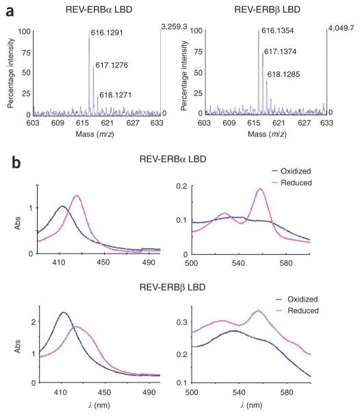
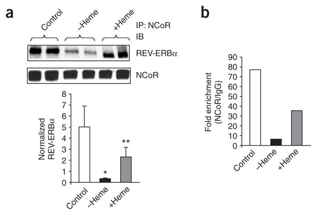
The nuclear receptors REV-ERBalpha (encoded by NR1D1) and REV-ERBbeta (NR1D2) have remained orphans owing to the lack of identified physiological ligands. Here we show that heme is a physiological ligand of both receptors. Heme associates with the ligand-binding domains of the REV-ERB receptors with a 1:1 stoichiometry and enhances the thermal stability of the proteins. Results from experiments of heme depletion in mammalian cells indicate that heme binding to REV-ERB causes the recruitment of the co-repressor NCoR, leading to repression of target genes including BMAL1 (official symbol ARNTL), an essential component of the circadian oscillator. Heme extends the known types of ligands used by the human nuclear receptor family beyond the endocrine hormones and dietary lipids described so far. Our results further indicate that heme regulation of REV-ERBs may link the control of metabolism and the mammalian clock.


Figures

Figure 1
Figure 1
Association of heme with the LBDs from REV-ERBα and REVERBβ. (a) MALDI mass spectra showing iron protoporphyrin IX in purified REV-ERBα and REV-ERBβ LBDs prepared from E. coli. Signals at 616 ± 2 Da correspond to heme iron-protoporphyrin IX. (b) Visible absorption spectra of REV-ERB LBD proteins. Peaks characteristic of heme–protein complexes are shown in the 390–450 nm range for the Soret band and in the 500–580 nm range for the α and β bands. Red spectra correspond to the heme–protein complexes after dithionite reduction of the iron moiety.
Figure 2
Figure 2
Thermodynamics of heme association with the REV-ERB LBDs measured by ITC and circular dichroism spectroscopy. (a,b) ITC data corresponding to the REV-ERBα (a) and REV-ERBβ (b) LBDs binding hemin. (c,d) Far-ultraviolet circular dichroism thermal melts corresponding to the REV-ERBα (c) and REV-ERBβ (d) LBDs in the heme-bound (+heme) and apoprotein (−heme) forms. The Tm values are 48.6 °C (−heme) and 52.9 °C (+heme) for REV-ERBα LBD and 48.4 °C (−heme) and 53.3 °C (+heme) for REV-ERBβ LBD.
Figure 3
Figure 3
Role of heme in regulation of REV-ERBα LBD activity. (a) Ultraviolet-visible spectrum of wild-type REV-ERBα (blue) and H602F REV-ERBα (red) LDBs. The absorbance peaks at 280 nm indicate that similar amounts of wild-type and H602F protein were examined, whereas the loss of the absorbance peak at ~420 nm for the H602F protein indicates lack of heme binding. (b) Lack of heme binding by the H602F REV-ERBα LBD mutant determined by ITC. Minimal binding to heme was not saturable; thus, Kd could not be estimated (c) Co-transfection reporter assay, comparing the transcriptional activity of the wild-type and H602F REV-ERBα LBDs fused to the Gal4 DBD in HuH7 hepatoma cells and HEK293 cells. The cells were co-transfected with a vector containing five copies of the Gal4 UAS upstream of luciferase (5×UAS-SV40 pGL3 firefly luciferase reporter vector). The wild-type and H602F chimeric proteins were expressed in comparable amounts, as determined by immunoblotting (Supplementary Fig. 8). Experiments were performed in triplicate a minimum of three times, and the mean ± s.d. of a representative experiment is shown.
Figure 4
Figure 4
Effect of modulation of intracellular heme on expression of REV-ERB target genes in HepG2 cells. (a) Treatment of HepG2 cells with succinylacetone (−heme) results in depletion of intracellular heme, whereas addition of hemin results in an increase in intracellular heme (+heme). (b) Expression of ALAS1 increases in response to a decrease in intracellular heme, and decreases in response to an increase in intracellular heme. (c,d) Expression of BMAL1 (c) and ELOVL3 (d) increases with heme depletion and decreases with increased intracellular heme; both genes are known to be repressed by REV-ERBα. Data are the mean ± s.d. of triplicate wells. *P < 0.05.
Figure 5
Figure 5
Effect of intracellular heme on NCoR interaction with REV-ERBα and recruitment to promoters. (a) The interaction of REV-ERBα with NCoR in HepG2 cells was determined by coimmunoprecipitation. Heme was depleted by succinylacetone treatment (−heme), and added back (+heme) by supplying hemin to heme-depleted cells. Cellular extracts were immunoprecipitated with antibody to NCoR, and samples were analyzed by immunoblotting with an antibody to REV-ERBα. Experiments were performed a minimum of three times and a representative gel is shown (top). Use of IgG did not result in detectable REV-ERBα (Supplementary Fig. 7b). Histogram shows quantification of the REV-ERBα immunoblots (bottom). Data are the mean ± s.d. from three individual experiments. Student’s t-test: *P < 0.05 versus control, **P < 0.05 versus heme depletion. (b) Effect of intracellular heme on NCoR occupancy in the BMAL1 promoter in HepG2 cells. Heme was depleted by succinylacetone treatment (−heme), and added back by hemin supplementation to heme-depleted cells (+heme). Chromatin was immunoprecipitated with an antibody to NCoR, and DNA was quantified by quantitative PCR. A control using rabbit IgG was used for normalization. Histogram shows the results from a representative of three independent experiments.

References

    1. Miyajima N, et al. Two erbA homologs encoding proteins with different T3 binding capacities are transcribed from opposite DNA strands of the same genetic locus. Cell. 1989;57:31–39. - PubMed
    1. Miyajima N, et al. Identification of two novel members of erbA superfamily by molecular cloning: the gene products of the two are highly related to each other. Nucleic Acids Res. 1988;16:11057–11074. - PMC - PubMed
    1. Lazar MA, Hodin RA, Darling DS, Chin WW. A novel member of the thyroid/steroid hormone receptor family is encoded by the opposite strand of the rat c-erbAα transcriptional unit. Mol Cell Biol. 1989;9:1128–1136. - PMC - PubMed
    1. Bonnelye E, et al. Rev-erbβ, a new member of the nuclear receptor superfamily, is expressed in the nervous system during chicken development. Cell Growth Differ. 1994;5:1357–1365. - PubMed
    1. Dumas B, et al. A new orphan member of the nuclear hormone receptor superfamily closely related to Rev-Erb. Mol Endocrinol. 1994;8:996–1005. - PubMed

Publication types

MeSH terms