. 2007 Nov;3(11):e173.
doi: 10.1371/journal.ppat.0030173.

A model of bacterial intestinal infections in Drosophila melanogaster

Affiliations

A model of bacterial intestinal infections in Drosophila melanogaster

Nadine T Nehme et al. PLoS Pathog. 2007 Nov.

Abstract

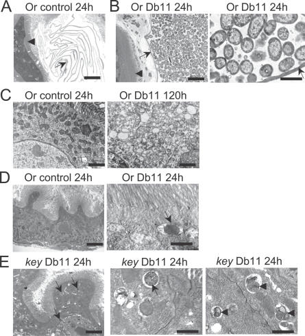
Serratia marcescens is an entomopathogenic bacterium that opportunistically infects a wide range of hosts, including humans. In a model of septic injury, if directly introduced into the body cavity of Drosophila, this pathogen is insensitive to the host's systemic immune response and kills flies in a day. We find that S. marcescens resistance to the Drosophila immune deficiency (imd)-mediated humoral response requires the bacterial lipopolysaccharide O-antigen. If ingested by Drosophila, bacteria cross the gut and penetrate the body cavity. During this passage, the bacteria can be observed within the cells of the intestinal epithelium. In such an oral infection model, the flies succumb to infection only after 6 days. We demonstrate that two complementary host defense mechanisms act together against such food-borne infection: an antimicrobial response in the intestine that is regulated by the imd pathway and phagocytosis by hemocytes of bacteria that have escaped into the hemolymph. Interestingly, bacteria present in the hemolymph elicit a systemic immune response only when phagocytosis is blocked. Our observations support a model wherein peptidoglycan fragments released during bacterial growth activate the imd pathway and do not back a proposed role for phagocytosis in the immune activation of the fat body. Thanks to the genetic tools available in both host and pathogen, the molecular dissection of the interactions between S. marcescens and Drosophila will provide a useful paradigm for deciphering intestinal pathogenesis.


Conflict of interest statement

Competing interests. The authors have declared that no competing interests exist.

Figures

Figure 1
Figure 1. S. marcescens Is Resistant to the Humoral Immune Response in the Septic Injury Model
(A–C) Wild-type (wt), Dif, or key flies were pricked using a thin needle dipped in a diluted overnight culture of Db11 (A), 20C2 (B), or Db1140 (C) (OD600 = 0.1) to introduce about 100 bacteria per fly. Flies were kept at 25 °C. Survival was monitored and expressed in % of surviving flies. Note that 20C2 and Db1140 kill key flies while they are less virulent in wild-type or Dif flies. (D) The hemolymph of pricked flies incubated was collected and spread on agar plates containing the appropriate antibiotic. The experiment was performed at 20 °C to insure that we would obtain enough surviving flies at the late time points of the experiment. 20C2 bacteria are kept in check in the hemolymph of wild-type, but not key flies. CFU: colony-forming units (logarithmic scale). (E) Flies were either preinjected with latex beads (LXB) or nontreated and then submitted to an immune challenge with Db11, except for nonchallenged controls. Survival was monitored at 20 °C. (F) Diptericin expression was induced in wild-type flies at 20 °C after pricking with Db11 or 20C2 but not in key mutant flies. Diptericin mRNA levels were measured by quantitative RT-PCR. The expression induced 24 h after an E. coli septic injury is taken as a reference. Dif flies exhibited normal induction of Diptericin after S. marcescens septic injury (not shown).
Figure 2
Figure 2. Ingested S. marcescens Db11 Kills Drosophila Flies Slowly
(A–C) Db11 (A) or 20C2 (B) bacteria were fed to wild-type (wt) or mutant flies at 25 °C. Survival is expressed in % of surviving flies. (A) median survival times in days (LT50): wt: 5.7; Dif: 5.0; key: 4.0; Dif. key: 3.0 (the difference between wt and all mutants is significant (p < 0.0001)); (B) wt: 6.7; Dif: 6.7; key: 4.5; Dif, key: 4.0 (the difference between wt and key is significant (p < 0.0001)). LPS-defective 20C2 kills wild-type flies more slowly than Db11: LT50 for flies feeding on 20C2 is 6.7 d compared to 5.7 d for flies on Db11 (p < 0.0001). (C) Flies were fed on a 50-mM sugar solution as control. All three experiments were performed in parallel. (D) The hemolymph of batches of 20 infected flies was collected and plated on LB agar containing the appropriate antibiotics. CFU: colony-forming units (logarithmic scale). The values for the 72-h time point are as follows: wt-Db11-GFP: 4.0 +/-0.26 (standard deviation); key-Db11-GFP: 5.1 +/-0.21; wt-20C2-GFP: 1.18 +/- 0.8 ; key-20C2-GFP: 4.4 +/- 0.3 (n = 4 experiments). (E) Guts were dissected and crushed, and dilutions of the extracts were plated as in (D). Note that flies are continuously feeding on a bacteria-containing sucrose solution. (F) Summary of the properties of the S. marcescens strains used in this study. Vir: virulent; att: attenuated virulence.
Figure 3
Figure 3. Effect of S. marcescens Oral Infection on the Structure of the Midgut
(A) S. marcescens Db11-GFP is found throughout the digestive tract after ingestion in both wild-type (upper panels) and key flies (lower panels), as observed with a dissecting microscope equipped with epifluorescent illumination. The fluorescent image (FITC channel) has been superimposed onto an oblique transmitted-light brightfield picture (except for the top left panel). Anterior is to the left. The cardia (proventriculus) is indicated by an asterisk. Scale bar is 150 μm. (B) The bacteria distend the gut lumen (indicated by arrowheads) during the later stages of the infection. Note the thinning of the epithelium in places where the digestive tract is bloated. Scale bar is 150 μm. Intestines of insects feeding on Db11 for 24 h are indistinguishable from those of control insects fed on sugar solution. (C–G) Confocal optical sections from the anterior part of the midgut of insects feeding on Db11-DsRed, after fixation and FITC-labeled phalloidin staining, that reveals the actin cytoskeleton enriched in the apical region of gut cells. Left panels show a longitudinal section and right panels show the transverse section from the axis displayed in each left panel. Note the thinning of the intestinal epithelium after 72 h (D) or 96 h (E) of infection as compared to the control feeding on sucrose (F) or after 24 h (C) of infection (OD = 0.1). (C–G) Scale bars are 50 μm. (G) Db11-DsRed infection at higher concentration (OD = 0.5) observed after 48 h: a bacterium inside a cell is shown at the center of the main panel and is indicated by circles in the lateral sections. (H) Immunostainings on frozen sections from the midgut of wild-type Oregon (Or) or key mutant flies fed for 6 h on sucrose (NI: noninfected) or OD = 0.2 Db11-DsRed (Inf: infected). The α-spectrin antibody staining (green) is enriched at the cell periphery. Nucleic acids of the epithelial cell and bacteria (arrows) is shown in red (DAPI staining). Scale bar is 10 μm.
Figure 4
Figure 4. S. marcescens Is Found within Intracellular Vacuoles in the Midgut Epithelium
Ultrastructure of wild-type and key mutant midguts fed on Db11 (OD = 0.1) or sucrose. (A, B) The midgut epithelium with associated microvilli (arrowhead) is separated from the gut lumen by the peritrophic membrane (arrow). (A) Oregon fly fed for 24 h on sucrose. Note that the peritrophic membrane is sometimes folded extensively and that no microorganisms are present. (B) Oregon fly fed for 24 h on Db11. The bacteria are confined inside of the peritrophic membrane and are shown at high resolution in the right panel. Scale bars: (A, B) left panel: 5 μm; (B) right panel: 1 μm. (C) Oregon fly fed for 24 h on sucrose (left panel) or for 120 h on Db11 (right panel). The infected midgut epithelial cell displays translucent vacuoles evocative of cellular stress. (D) Oregon fly fed for 24 h on sucrose (left panel) or for 24 h on Db11 (right panel). An intracellular bacterium inside a vacuole is indicated by an arrow. (E) key mutant fly fed for 24 h on Db11. Intracellular bacteria are indicated by arrows and putative degraded bacteria by arrowheads. Scale bars are 5 μm for low magnification pictures ([A], [C], left panel in [B], [D], and [E]) and 1 μm in high magnification pictures (right panels in [B], [D], and [E]).
Figure 5
Figure 5. The Systemic Humoral Immune Response Is Not Triggered by Ingested S. marcescens
(A) Diptericin mRNA levels were measured by quantitative RT-PCR. No significant expression was induced after feeding with S. marcescens Db11 or 20C2, or with nonpathogenic E. coli 1106 (wt, Dif, key, and wt E. coli samples, respectively). The expression induced by an E. coli 1106 septic injury is taken as a reference (*). No induction of Cecropin expression could be detected; the systemic immune response was not significantly induced at later time points (up to 144 h; unpublished data). (B) No expression of ß-galactosidase was detected in fat body cells of Diptericin-lacZ transgenic flies as indicated by the absence of blue staining in fat body lobules (arrowheads). The expression observed in wild-type flies is in pericardial cells and is also found in key mutants, although they are absent in this dissection. (C) Flies that have fed on Db11 for 48 h (primary challenge) are still able to mount a systemic immune response when pricked by either E. coli (secondary challenge: 6 h after injury) or Db11 (not shown). This response is dependent on the imd pathway as key mutants showed no response. Diptericin mRNA levels were measured by quantitative RT-PCR 54 h after the beginning of bacterial ingestion. *: 100 % reference: wild-type flies challenged with E. coli for 6 h. (D) Flies fed on Db11 are still able to mount an effective immune response since they do not succumb to a secondary E. coli 1106 septic injury (wt and key (E. coli)) at an enhanced rate, as compared to mock-challenged flies (wt and key (ci); ci: clean injury). The arrow indicates the time point (day 2) at which the secondary challenge was performed (all curves correspond to flies that were feeding on Db11, except for the solid yellow and green lines, which correspond respectively to control wt and key flies fed on sugar [S] solution and challenged with E. coli at day 2). wt and key flies feeding on Db11 and not challenged secondarily by E. coli are also shown (wt and key).
Figure 6
Figure 6. The imd-Dependent Immune Response in the Midgut Mediates Host Defense against S. marcescens Intestinal Infections
(A) A Diptericin-GFP transgene is induced in the posterior part of the midgut following an oral challenge with S. marcescens Db11-DsRed (OD = 0, 1, 72 h), as observed by confocal microscopy. Scale bar is 50 μm. (B–E) The expression of Diptericin in the midgut was visualized in dissected preparations of flies carrying a Diptericin-lacZ reporter transgene that have fed on Db11 (B). This expression is reduced in the imd-pathway mutants PGRP-LC (C) and absent in key (D) midguts. The transgene is not induced in flies feeding on sugar solution (E). Scale bar is 150 μm. (F) key mutant flies expressing a Diptericin transgene in the midgut under the control of the NP3084 Gal4 driver (NP3084-Gal4, key/key, UAS-Diptericin) survive longer than key mutants following an oral infection at 29 °C with Db11 (median survival time [LT50]: 3.7 versus 3.0; p < 0.0001). The rescue of the key mutant phenotype by the expression of a key transgene (NP3084-Gal4, key/key; UAS-key) in the midgut is complete (LT50: 5.1, p < 0.0001). Similar results have been obtained with two other Gal4 drivers that induce the expression of UAS-transgenes in the midgut. Experiments have been repeated twice. NP3084-Gal4, key/key flies die at the same rate as key mutant flies (LT50 2.7 versus 3.0, p = 0.47). (G) Levels of Diptericin transcripts in midgut of wild-type and transgenic flies as measured by quantitative real-time RT-PCR. The normalized signal (using ribosomal protein 49 gene transcript) obtained from extracts of whole flies undergoing a systemic immune response has been arbitrarily set at 100%. The measured values are displayed on the top of each bar. The overexpression of Diptericin induced by the UAS-Dipt transgene (32% and 43% for flies fed with sugar and Db11 [Db11] or sugar alone [sugar]) is much higher than that observed in wild-type flies infected per os (3%).
Figure 7
Figure 7. Phagocytosis Plays an Essential Role in the Control of Intestinal S. marcescens Infection
(A) A pTEP1-GFP-expressing hemocyte that has phagocytosed Db11-DsRed bacteria. The picture was taken with the apotome microscope: left-panel (GFP); central panel (DsRed); right panel (merge). pTEP1-GFP expression specifically labels hemocytes. Bacteria that have been engulfed appear yellow in the right panel. (B–D) Phagocytosis inactivation through the injection of latex beads (+LXB) leads to the earlier demise of treated flies as compared to control (untreated flies). (B) Db11: median survival time (LT50 in days), wt: 5.63, wt+LXB: 2.63, key: 3.63, key +LXB: 1.92 (LXB-treated versus untreated flies: p < 0.0001; wt+LXB versus key +LXB: p = 0.0089). (C) 20C2: wt: 6.88, wt+LXB: 2.63, key: 3.63, key +LXB: 1.292 (LXB-treated versus untreated flies: p < 0.0001; wt+LXB versus key +LXB: p = 0.0045). Db1140 (D). Latex bead–injected flies usually survive the procedure well when fed on sucrose alone (not shown). (E–G) The hemolymph of latex bead-injected and nontreated surviving flies (batches of 20 flies) that had been feeding on Db11-GFP (E), 20C2-GFP (F), Db1140-GFP (G) was collected and plated on agar plates containing the appropriate antibiotics. The counts were performed only up to 48 h in the case of LXB-treated flies since most were dead by 72 h. CFU: colony-forming units (log scale). (H) Diptericin mRNA levels were measured by quantitative RT-PCR in whole flies after Db11 feeding or LXB injection and Db11 feeding. A strong expression was induced only after phagocytosis inactivation through the prior injection of LXB. The expression induced by an E. coli 1106 septic injury is taken as a reference.
Figure 8
Figure 8. The Increasing Number of Db11 in the Hemolymph Results from Bacterial Passage from the Gut
(A, B) Flies were fed for 24 h on Db11-GFP bacteria and then transferred to a vial containing only Db11-DsRed bacteria. The number of GFP- and DsRed-expressing bacteria was counted in the intestine (A) and in the hemolymph of latex bead–injected and nontreated surviving flies (batches of 20 flies) (B). The counts were performed only up to 48 h in the case of LXB-treated flies since most were dead by 72 h. CFU: colony-forming units (log scale).

References

    1. Royet J, Dziarski R. Peptidoglycan recognition proteins: pleiotropic sensors and effectors of antimicrobial defences. Nat Rev Microbiol. 2007;5:264–277. - PubMed
    1. Lemaitre B, Hoffmann J. The host defense of Drosophila melanogaster. Annu Rev Immunol. 2007;25:697–743. - PubMed
    1. Bulet P, Hetru C, Dimarcq JL, Hoffmann D. Antimicrobial peptides in insects; structure and function. Dev Comp Immunol. 1999;23:329–344. - PubMed
    1. Tzou P, Reichhart JM, Lemaitre B. Constitutive expression of a single antimicrobial peptide can restore wild-type resistance to infection in immunodeficient Drosophila mutants. Proc Natl Acad Sci U S A. 2002;99:2152–2157. - PMC - PubMed
    1. Michel T, Reichhart J, Hoffmann JA, Royet J. Drosophila Toll is activated by Gram-positive bacteria through a circulating peptidoglycan recognition protein. Nature. 2001;414:756–759. - PubMed

Publication types

MeSH terms

LinkOut - more resources