. 2007 Nov;3(11):e212.
doi: 10.1371/journal.pgen.0030212.

Extragenic accumulation of RNA polymerase II enhances transcription by RNA polymerase III

Affiliations

Extragenic accumulation of RNA polymerase II enhances transcription by RNA polymerase III

Imke Listerman et al. PLoS Genet. 2007 Nov.

Abstract

Recent genomic data indicate that RNA polymerase II (Pol II) function extends beyond conventional transcription of primarily protein-coding genes. Among the five snRNAs required for pre-mRNA splicing, only the U6 snRNA is synthesized by RNA polymerase III (Pol III). Here we address the question of how Pol II coordinates the expression of spliceosome components, including U6. We used chromatin immunoprecipitation (ChIP) and high-resolution mapping by PCR to localize both Pol II and Pol III to snRNA gene regions. We report the surprising finding that Pol II is highly concentrated approximately 300 bp upstream of all five active human U6 genes in vivo. The U6 snRNA, an essential component of the spliceosome, is synthesized by Pol III, whereas all other spliceosomal snRNAs are Pol II transcripts. Accordingly, U6 transcripts were terminated in a Pol III-specific manner, and Pol III localized to the transcribed gene regions. However, synthesis of both U6 and U2 snRNAs was alpha-amanitin-sensitive, indicating a requirement for Pol II activity in the expression of both snRNAs. Moreover, both Pol II and histone tail acetylation marks were lost from U6 promoters upon alpha-amanitin treatment. The results indicate that Pol II is concentrated at specific genomic regions from which it can regulate Pol III activity by a general mechanism. Consequently, Pol II coordinates expression of all RNA and protein components of the spliceosome.


Conflict of interest statement

Competing interests. The authors have declared that no competing interests exist.

Figures
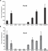
Figure 1
Figure 1. Both Pol II and Pol III Accumulate at Active U6 snRNA Gene Promoters
Sheared chromatin from pemHeLa cells that had been crosslinked with formaldehyde was immunoprecipitated with (A) anti-Pol III (1900) or (B) anti-Pol II antibodies (8WG16). DNA was purified following reversal of crosslinks. qPCR primers specific for regions ∼ 300 bp upstream of active and inactive U6 genes (U6–1, U6–2, U6–7, U6–8, U6–9 and U6–3, U6–4, U6–6, respectively), PGK1, and tRNALeu genes (Pol II and Pol III controls, respectively). All values are relative to nonimmune IgG and normalized to an intergenic control region. Error bars represent the SEM of at least three independent experiments.
Figure 2
Figure 2. Pol II and Pol III Accumulate in Two Distinct Peaks at the U6–1, U6–7, and U6–8 Promoters
(A, B) Diagrams of the U6–1 and U6–7/U6–8 gene regions, with black lines specifying the PCR amplicons identified by the central nucleotide. Boxes represent the position of the DSE. (C, D) Summary plots of ChIP experiments with Pol II and Pol III distributions on U6–1 gene region (C) and U6–7 and U6–8 (D). Crosslinked pemHeLa extracts were used for ChIP with primers amplifying fragments distributed 1 kb upstream and downstream of each gene. Antibodies specific for Pol II (4H8) and Pol III were used. X-axis values in (C) are relative to the U6–1 transcription start site at position +1, whereas transcription start sites in (D) are at +1,302 (U6–7) and +2,426 (U6–8), relative to an arbitrary point upstream of U6–7. Data points are placed according to the center positions of the PCR products along the region. The peak value for the region (either U6–1 region or U6–7/U6–8 region) was determined for each biological replicate and set to 100%; displayed values are averaged among at least three biological replicates. Error bars represent the SEM.
Figure 3
Figure 3. Comparison of Pol II Accumulation Pattern on U6–1 and U1 snRNA Genes
ChIP with Pol II mAb 4H8 was performed with pemHeLa crosslinked extracts and the purified DNA subjected to qPCR with primers along the U6–1 and U1 snRNA gene regions as shown (Ensembl ID ENSG00000194297 on chromosome 1). Data points represent the center point of the amplified region and Pol II fold accumulation over background and intergenic region, as in Figure 1. Diagrams below the plot indicate the features of both genes, which have been aligned at their respective transcription start sites (arrows). Error bars represent the SEM of at least three independent experiments.
Figure 4
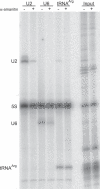
Figure 4. α-Amanitin Reduces Endogenous U6 snRNA Expression
HeLa cells were transfected with a tRNAArg maxigene and 16 h later treated with 10 μg/ml α-amanitin or left untreated. After 3 h, 250 μCi [32P] orthophosphate was added to the medium for 6 h. Total RNA was collected and 5S rRNA along with tRNAArg, U2 and U6 snRNAs were hybrid selected with biotinylated complementary oligos. Hybrid-selected RNA was separated on a denaturating polyacrylamide gel and exposed to PhosphorImager plates. The 5S rRNA bands served as a loading control. Three biological replicates were analyzed and a representative gel is shown.
Figure 5
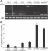
Figure 5. Low α-Amanitin Concentrations Inhibit U6 snRNA Maxigene Expression
HeLa cells were transiently transfected with 1 μg U6–1, U6–8, and U6–9 maxigenes carrying a 9 bp insertion and treated simultaneously with 50 nM α-amanitin oleate for 20 h or left untreated. Expression of U6 maxigenes, LDHA, 28S rRNA, 5S rRNA and pre-tRNATyr was measured by gene-specific reverse transcription, followed by conventional PCR and agarose gel electrophoresis (A) or qPCR (B). qPCR values were normalized to 28S rRNA and expression levels are expressed relative to untreated controls. Error bars represent the SEM. The data represents the average of at least three independent experiments.
Figure 6
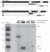
Figure 6. Pol III Transcribes U6–1 Maxigene
Primer extension analysis of RNA from HeLa cells cotransfected with GFP expression plasmid and U6–1 maxigene. (A) Design of the two U6–1 maxigenes with insertions at +66 and +87 bp (white boxes) to allow for maxigene-specific reverse transcription. Construct “U6–1 maxiT” harbors five thymidine residues directly downstream of the linker insertion; “U6–1 MaxiC” harbors the same primer binding site but lacks the T's. The cross-hatched box represents the reverse primer specific for the downstream insertion, yielding extension products of either 114 bp (U6–1 maxiT) or 109 bp (U6–1 maxiC). Grey filled box and black filled box represent reverse primers specific for upstream insertion with or without T residues, respectively, leading to extension products of 83 bp (U6–1 maxiT) or 79 bp (U6–1 maxiC). Primer extension of mRNA derived from cotransfected GFP plasmid yields a 158 bp product. Primer extension products were separated on a denaturing polyacrylamide gel and exposed on a PhosphorImager. (B) A representative gel is shown.
Figure 7
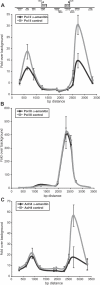
Figure 7. Pol II and Histone Acetylation Levels at U6 Genes Are Reduced by α-Amanitin
Extracts from crosslinked Hela cells, either treated for 9 h with 10 μg/ml α-amanitin or left untreated, were subjected to ChIP with (A) Pol II, (B) Pol III, or (C) acetylated histone H4 antibodies. Purified DNA was subjected to qPCR with primers along U6–7 and U6–8 gene regions. All values are relative to background and normalized to an intergenic control region. Error bars represent the SEM. The data represents the average of three to five independent experiments.

References

    1. Jurica MS, Moore MJ. Pre-mRNA splicing: awash in a sea of proteins. Mol Cell. 2003;12:5–14. - PubMed
    1. Brow DA, Guthrie C. Spliceosomal RNA U6 is remarkably conserved from yeast to mammals. 1988;334:213–218. - PubMed
    1. Vidal VP, Verdone L, Mayes AE, Beggs JD. Characterization of U6 snRNA-protein interactions. RNA. 1999;5:1470–1481. - PMC - PubMed
    1. Stanek D, Neugebauer KM. The Cajal body: a meeting place for spliceosomal snRNPs in the nuclear maze. Chromosoma. 2006;115:343–354. - PubMed
    1. Kunkel GR, Maser RL, Calvet JP, Pederson T. U6 small nuclear RNA is transcribed by RNA polymerase III. Proc Natl Acad Sci U S A. 1986;83:8575–8579. - PMC - PubMed

Publication types

MeSH terms