

The inflammatory response to cell death
- PMID: 18039143
- PMCID: PMC3094097
- DOI: 10.1146/annurev.pathmechdis.3.121806.151456
The inflammatory response to cell death
Abstract
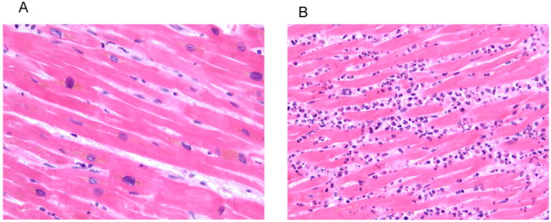
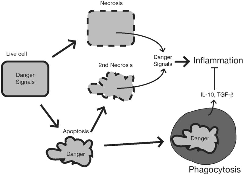
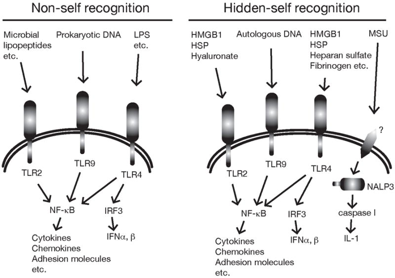
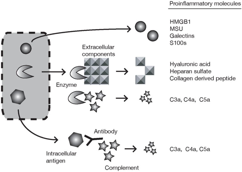
When cells die in vivo, they trigger an inflammatory response. The ensuing hyperemia, leak of plasma proteins, and recruitment of leukocytes serve a number of useful functions in host defense and tissue repair. However, this response can also cause tissue damage and contribute to the pathogenesis of a number of diseases. Given the key role of inflammation in these processes, it is important to understand the underlying mechanisms that drive this response. Injured cells release danger signals that alert the host to cell death. Some of these molecules are recognized by cellular receptors that stimulate the generation of proinflammatory mediators. Other molecules released by dead cells stimulate the generation of mediators from extracellular sources. The resulting mediators then orchestrate the inflammatory response, eliciting its various vascular and cellular components. Dead cells also release danger signals that activate dendritic cells and promote the generation of immune responses to antigens. Here we review what is presently known about the sterile inflammatory response and its underlying mechanisms.
Figures
References
-
- Steinhoff M, Vergnolle N, Young SH, Tognetto M, Amadesi S, Ennes HS, Trevisani M, Hollenberg MD, Wallace JL, Caughey GH, Mitchell SE, Williams LM, Geppetti P, Mayer EA, Bunnett NW. Agonists of proteinase-activated receptor 2 induce inflammation by a neurogenic mechanism. Nat Med. 2000;6:151–8. - PubMed
-
- Kaplan AP, Joseph K, Silverberg M. Pathways for bradykinin formation and inflammatory disease. J Allergy Clin Immunol. 2002;109:195–209. - PubMed
-
- Klinger MH. Platelets and inflammation. Anat Embryol (Berl) 1997;196:1–11. - PubMed
-
- Majno G, La Gattuta M, Thompson TE. Cellular death and necrosis: chemical, physical and morphologic changes in rat liver. Virchows Arch Pathol Anat Physiol Klin Med. 1960;333:421–65. - PubMed
Publication types
MeSH terms
Substances
Grants and funding
LinkOut - more resources
Full Text Sources
Other Literature Sources
