. 2007 Nov 26:4:28.
doi: 10.1186/1742-2094-4-28.

Developmental stage of oligodendrocytes determines their response to activated microglia in vitro

Affiliations

Developmental stage of oligodendrocytes determines their response to activated microglia in vitro

Brandon A Miller et al. J Neuroinflammation. .

Abstract

Background: Oligodendrocyte progenitor cells (OPCs) and mature oligodendrocytes are both lost in central nervous system injury and disease. Activated microglia may play a role in OPC and oligodendrocyte loss or replacement, but it is not clear how the responses of OPCs and oligodendrocytes to activated microglia differ.

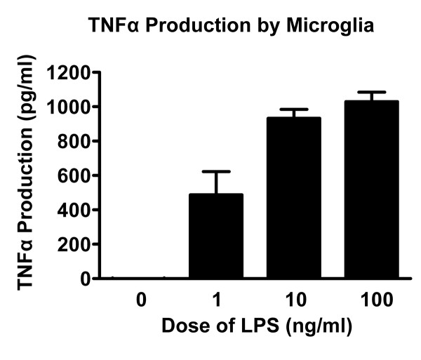
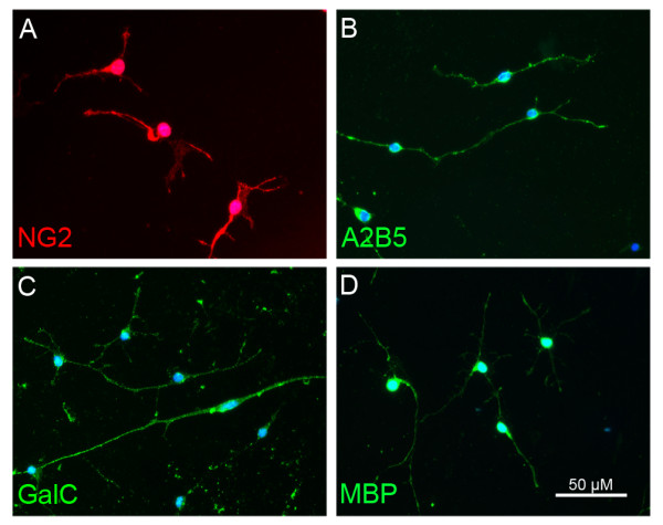
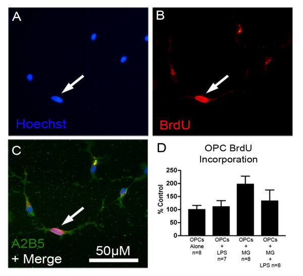
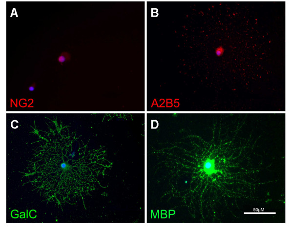
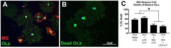
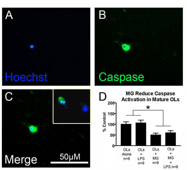
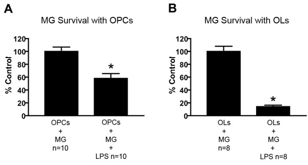
Methods: OPCs and microglia were isolated from rat cortex. OPCs were induced to differentiate into oligodendrocytes with thyroid hormone in defined medium. For selected experiments, microglia were added to OPC or oligodendrocyte cultures. Lipopolysaccharide was used to activate microglia and microglial activation was confirmed by TNFalpha ELISA. Cell survival was assessed with immunocytochemistry and cell counts. OPC proliferation and oligodendrocyte apoptosis were also assessed.

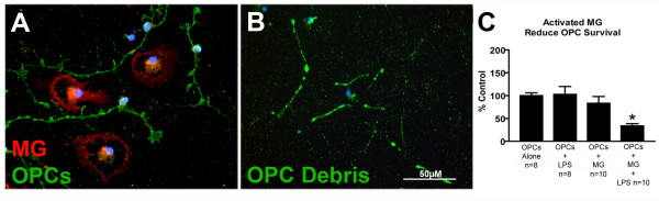
Results: OPCs and oligodendrocytes displayed phenotypes representative of immature and mature oligodendrocytes, respectively. Activated microglia reduced OPC survival, but increased survival and reduced apoptosis of mature oligodendrocytes. Activated microglia also underwent cell death themselves.

Conclusion: Activated microglia may have divergent effects on OPCs and mature oligodendrocytes, reducing OPC survival and increasing mature oligodendrocyte survival. This may be of importance because activated microglia are present in several disease states where both OPCs and mature oligodendrocytes are also reacting to injury. Activated microglia may simultaneously have deleterious and helpful effects on different cells after central nervous system injury.


Figures

Figure 1
Figure 1
TNFα production by microglia. In a preliminary experiment, microglia alone were treated with LPS at varying doses and TNFα production was assessed 24 hours later. LPS induced TNFα production in a dose dependent manner.
Figure 2
Figure 2
OPC morphology. OPCs grown in media containing bFGF/PDGF labeled for oligodendrocyte markers at day 3 in vitro. OPCs labeled positively for NG2 (A), A2B5 (B), GalC (C) and MBP (D).
Figure 3
Figure 3
LPS-activated microglia induce OPC cell death. OPCs were cultured in combination with microglia (A). Dead OPCs were not able to be quantified due to scattered debris (B), so OPC survival was measured by counting the number of live OPCs. LPS or microglia alone had no effect on OPC survival but microglia activated with LPS significantly reduced OPC survival (C, * = p < 0.05 from control, Tukey's, error bars = SEM).
Figure 4
Figure 4
OPC proliferation as measured by BrdU incorporation. To assure that reduced OPC survival in the presence of LPS-activated microglia was not due to a reduction in OPC proliferation, a BrdU incorporation study was conducted. Some OPC nuclei (A) also incorporated BrdU (B). OPCs were also labeled with antibody to A2B5 (C) to confirm that BrdU positive cells were OPCs. OPC proliferation, as assessed by cell counts, was not altered by microglia or LPS-activated microglia (though there was a trend toward increased OPC proliferation in the presence of microglia, p = 0.056, factorial ANOVA for main effect of microglia) verifying that lower OPC numbers were due to OPC loss rather than reduced OPC proliferation (D, error bars = SEM).
Figure 5
Figure 5
Oligodendrocyte morphology. Oligodendrocytes grown in media containing thyroid hormone labeled for oligodendrocyte markers at day 3 in vitro. Oligodendrocytes did not label positively for the immature OPC markers NG2 (A) or A2B5 (B), but did label for the mature oligodendrocyte markers GalC (C) and MBP (D).
Figure 6
Figure 6
Microglia and LPS-activated microglia both increase oligodendrocyte survival. Oligodendrocytes were cultured in combination with microglia (A). Dead oligodendrocytes were visible by cellular debris left behind after fixation and staining (B) and therefore oligodendrocyte cell death was quantified as percent cell death based on cell counts. Both microglia and microglia activated by LPS significantly reduced the percentage of dead oligodendrocytes (C, * = p < 0.05, factorial ANOVA for main effect of microglia, error bars = SEM). The protective effect of microglia was unchanged by the presence of LPS (p < 0.05 factorial ANOVA).
Figure 7
Figure 7
Microglia reduce caspase activation in oligodendrocytes. Fragmented and condensed nuclei in oligodendrocyte cultures (A) were found in all cell bodies that labeled with a caspase indicator probe (B), which allowed for identification of oligodendrocytes by morphology. Overlap of apoptotic-morphology nuclei and the caspase indicator were used to confirm apoptotic cell death (C). The inset in panel C shows another representative image of a fragmented nucleus in an oligodendrocyte positive for caspase activation. Cell counts were conducted to quantify caspase activation (D, * = p < 0.05, factorial ANOVA for main effect of microglia, error bars = SEM). Microglia and LPS-activated microglia both reduced oligodendrocyte apoptosis, suggesting that increased oligodendrocyte survival in the presence of microglia is due to a reduction in apoptosis. As in figure 5, the protective effect of microglia was unchanged by the presence of LPS (factorial ANOVA).
Figure 8
Figure 8
Assessment of microglial cell death. In experiments in which microglia were combined with OPCs, 10 ng/ml LPS significantly reduced microglia number to 58 ± 8% of control, based on cell counts (A). In experiments in which microglia were combined with oligodendrocytes, 10 ng/ml LPS significantly reduced microglia number to 14 ± 2% of control (B, * = p < 0.05, T-test, error bars = SEM).

References

    1. Raff MC, Miller RH, Noble M. A glial progenitor cell that develops in vitro into an astrocyte or an oligodendrocyte depending on culture medium. Nature. 1983;303:390–396. doi: 10.1038/303390a0. - DOI - PubMed
    1. Wolswijk G, Noble M. Identification of an adult-specific glial progenitor cell. Development. 1989;105:387–400. - PubMed
    1. Scolding NJ, Rayner PJ, Compston DA. Identification of A2B5-positive putative oligodendrocyte progenitor cells and A2B5-positive astrocytes in adult human white matter. Neuroscience. 1999;89:1–4. doi: 10.1016/S0306-4522(98)00548-X. - DOI - PubMed
    1. McTigue DM, Horner PJ, Stokes BT, Gage FH. Neurotrophin-3 and brain-derived neurotrophic factor induce oligodendrocyte proliferation and myelination of regenerating axons in the contused adult rat spinal cord. J Neurosci. 1998;18:5354–5365. - PMC - PubMed
    1. Sun F, Lin C, McTigue DM, Shan X, C.A. T, J.C. B, Beattie MS. Dorsal rhizotomy induces oligodendrocyte progenitor proliferation and oligodendrocyte genesis, while spinal contusion induces oligodendrocyte death. J Neurosci. 2006.

Publication types

MeSH terms