. 2008 Mar;29(3):494-500.
doi: 10.3174/ajnr.A0847. Epub 2007 Nov 26.

A T1 hyperintense perilesional signal aids in the differentiation of a cavernous angioma from other hemorrhagic masses

Affiliations

A T1 hyperintense perilesional signal aids in the differentiation of a cavernous angioma from other hemorrhagic masses

T J Yun et al. AJNR Am J Neuroradiol. 2008 Mar.

Abstract

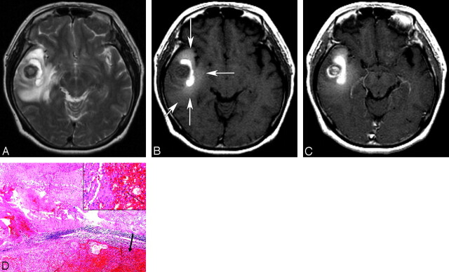
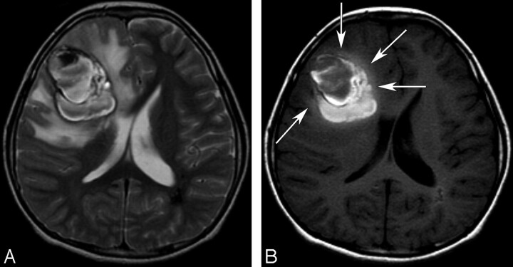
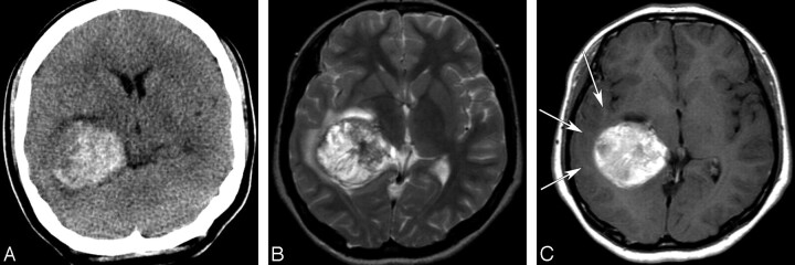
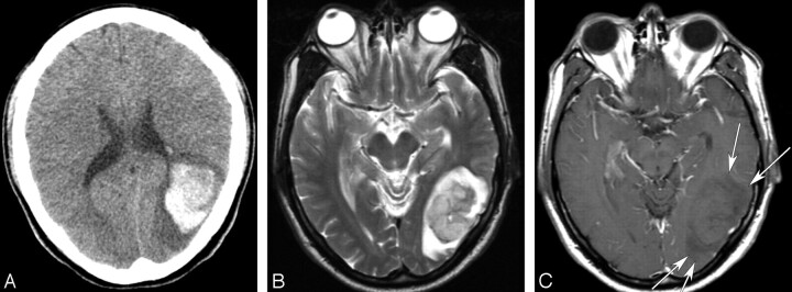
Background and purpose: A cavernous angioma is a developmental vascular malformation with a high risk of hemorrhage. The purpose of this work was to retrospectively determine whether an MR sign of T1 hyperintense perilesional signal intensity is useful for the differentiation of cavernous angioma from other hemorrhagic cerebral masses.

Materials and methods: The institutional review board approved this study. We retrospectively evaluated the MR images of 72 patients with acute or subacute cerebral hemorrhagic lesions with perilesional edema (29 cavernous angiomas, 13 glioblastomas, 1 oligodendroglioma, 16 metastatic tumors, and 13 intracerebral hemorrhages) for the presence of T1 hyperintense perilesional signal intensity. In addition, T1 signal intensities of a perilesional edema were quantitatively analyzed. In cavernous angiomas, volumes of hemorrhagic lesions and perilesional edemas, lesion locations, presence of contrast enhancement, and time intervals between symptom onset and MR imaging were also assessed. Data were analyzed using unpaired t test or Fisher exact test.

Results: T1 hyperintense perilesional signal intensity sign was found in 18 (62.1%) of 29 cavernous angiomas, in 1 (6.3%) of 16 metastases, and in 0 primary brain tumors or intracerebral hemorrhages. Sensitivity, specificity, and positive predictive value of this sign for cavernous angioma were 62%, 98%, and 95%, respectively. The perilesional T1 hyperintensity was significantly higher in cavernous angiomas (P = .045) than in normal white matter. Perilesional edema volumes were larger in cavernous angiomas with the MR sign than in cavernous angiomas without the sign (P = .009).

Conclusion: When the MR sign of T1 hyperintense perilesional signal intensity is present, there is a high probability of cavernous angioma being present in the brain, and this MR sign may be helpful for differentiating cavernous angioma from hemorrhagic tumors and intracerebral hemorrhages.


Figures

Fig 1.
Fig 1.
A 54-year-old woman with a cavernous angioma. A, An axial T2-weighted image (TR/TE/NEX, 3700/104/2 ms) shows a hemorrhagic mass with heterogeneous signal intensity and a severe surrounding edema in the right temporal lobe. B, An unenhanced T1-weighted image (TR/TE/NEX, 467/8/1 ms) demonstrates obvious hyperintensity within the vasogenic edema (T1 hyperintense perilesional signal intensity sign; long arrows). The T1 hyperintense perilesional signal intensity sign is not obviously observed in the peripheral area of the vasogenic edema (short arrow). C, An enhanced T1-weighted image (TR/TE/NEX, 467/8/1 ms) shows heterogeneous enhancement of the round mass at the center of hemorrhagic mass. D, Surgery was performed 1 day after MR imaging. A histologic photomicrograph shows a relatively well-demarcated cavernous angioma with hematoma formation (arrow) and microscopic diffuse perilesional hemorrhage in the surrounding white matter (hematoxylin-eosin, original magnification, ×40). The inset shows hemorrhage with infiltration of siderophages (arrow; hematoxylin-eosin, original magnification, ×200).
Fig 2.
Fig 2.
An 11-year-old boy with a cavernous angioma. A, An axial T2-weighted image (TR/TE/NEX, 3683/104/2 ms) shows a large hemorrhagic mass with heterogeneous signal intensity and peripheral hypointense rims in the right frontal lobe. A perilesional massive edema and mass effect is seen. There is a hypointense lesion in the periventricular white matter of the left parietal lobe and a few small hypointense lesions that were also found in both hemispheres on a T2* gradient-echo image (data not shown), which indicate possible multiple cavernous angiomas. B, An axial T1-weighted image (TR/TE/NEX, 500/9/2 ms) shows a T1 hyperintense perilesional signal intensity sign and mild hyperintensity of a perilesional edema at the deep area abutting the hemorrhagic mass (arrows). Note the centripetal pattern of the T1 hyperintense perilesional signal intensity sign in which T1 hyperintensity within the perilesional edema is observed only in the deep area around the hemorrhagic mass; it is not observed at the periphery of edema.
Fig 3.
Fig 3.
A 17-year-old girl with a glioblastoma. A, An unenhanced CT image shows a hyperattenuated acute hemorrhagic mass in the right temporal lobe. B, An axial T2-weighted image (TR/TE/NEX, 5000/96/2 ms) shows a hemorrhagic mass with heterogeneous signal intensity and a mild peritumoral edema. C, An axial enhanced T1-weighted image (TR/TE/NEX, 500/12/1 ms) demonstrates mild hypointensity or isointensity of the peritumoral edema (arrows) and mild heterogeneous enhancement of the hemorrhagic mass, which was hyperintense on an unenhanced T1-weighted image (data not shown).
Fig 4.
Fig 4.
A 48-year-old man with a cerebral metastasis from a hepatocellular carcinoma. A, An unenhanced CT image shows an acute hemorrhagic mass with a surrounding edema in the left temporooccipital lobe. B, An axial T2-weighted image (TR/TE/NEX, 5000/99/2 ms) demonstrates an acute hematoma with a profound perilesional edema and mass effect. C, An axial enhanced T1-weighted image (TR/TE/NEX, 500/12/1 ms) demonstrates hypointensity of the perilesional edema (arrows).

References

    1. Zabramski JM, Wascher TM, Spetzler RF, et al. The natural history of familial cavernous malformations: results of an ongoing study. J Neurosurg 1994;80:422–32 - PubMed
    1. Rigamonti D, Drayer BP, Johnson PC, et al. The MRI appearance of cavernous malformations (angiomas). J Neurosurg 1987;67:518–24 - PubMed
    1. Dorsch NWC, McMahon JHA. Intracranial cavernous malformations—natural history and management. Crit Rev Neurosurg 1998;8:154–68 - PubMed
    1. Moran NF, Fish DR, Kitchen N, et al. Supratentorial cavernous haemangiomas and epilepsy: a review of the literature and case series. J Neurol Neurosurg Psychiatry 1999;66:561–68 - PMC - PubMed
    1. Sandalcioglu IE, Wiedemayer H, Secer S, et al. Surgical removal of brain stem cavernous malformations: surgical indications, technical considerations, and results. J Neurol Neurosurg Psychiatry 2002;72:351–55 - PMC - PubMed