. 2007 Nov;177(3):1377-94.
doi: 10.1534/genetics.107.070425.

Phylogenetic footprinting analysis in the upstream regulatory regions of the Drosophila enhancer of split genes

Affiliations

Phylogenetic footprinting analysis in the upstream regulatory regions of the Drosophila enhancer of split genes

Morgan L Maeder et al. Genetics. 2007 Nov.

Abstract

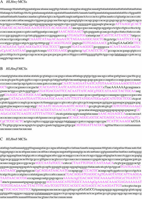
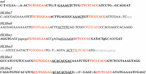
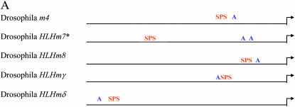
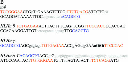
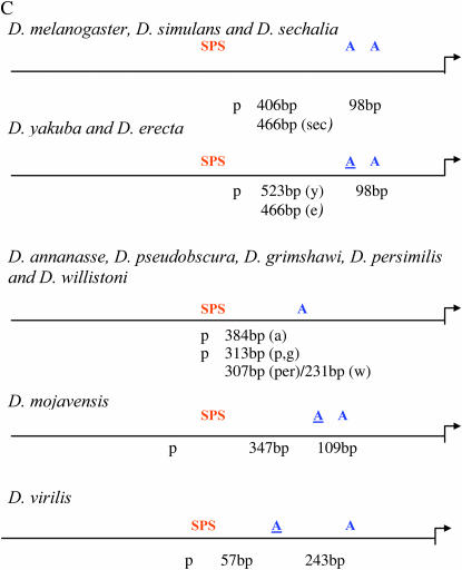
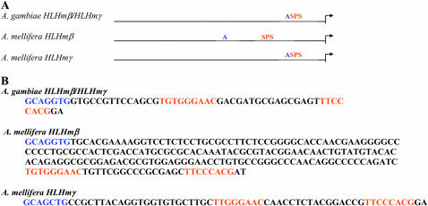
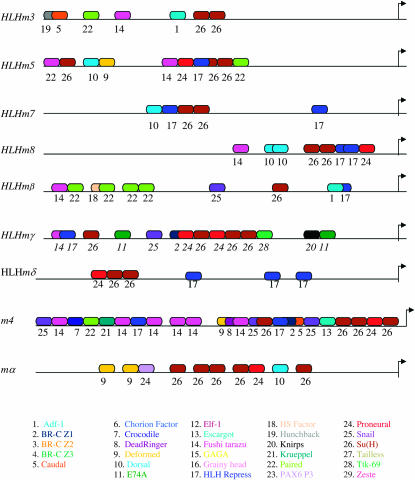
During Drosophila development Suppressor of Hairless [Su(H)]-dependent Notch activation upregulates transcription of the Enhancer of split-Complex [E(spl)-C] genes. Drosophila melanogaster E(spl) genes share common transcription regulators including binding sites for Su(H), proneural, and E(spl) basic-helix-loop-helix (bHLH) proteins. However, the expression patterns of E(spl) genes during development suggest that additional factors are involved. To better understand regulators responsible for these expression patterns, recently available sequence and annotation data for multiple Drosophila genomes were used to compare the E(spl) upstream regulatory regions from more than nine Drosophila species. The mgamma and mbeta regulatory regions are the most conserved of the bHLH genes. Fine analysis of Su(H) sites showed that high-affinity Su(H) paired sites and the Su(H) paired site plus proneural site (SPS + A) architecture are completely conserved in a subset of Drosophila E(spl) genes. The SPS + A module is also present in the upstream regulatory regions of the more ancient mosquito and honeybee E(spl) bHLH genes. Additional transcription factor binding sites were identified upstream of the E(spl) genes and compared between species of Drosophila. Conserved sites provide new understandings about E(spl) regulation during development. Conserved novel sequences found upstream of multiple E(spl) genes may play a role in the expression of these genes.


Figures

F<sc>igure</sc> 1.—
Figure 1.—
Identification of multispecies conserved sequences in the promoters of E(spl) HLHmγ, HLHmβ, and HLHm8. EvoPrinter outputs for HLHmγ (A), HLHmβ (B), and HLHm8 (C) are shown. Uppercase letters in pink represent nucleotides in the 1200-bp D. melanogaster reference multispecies conserved sequences (MCSs) that are conserved in D. simulans, D. yakuba, D. erecta, D. annanasse, D. pseudoobscura, D. virilis, D. mojavensis, and D. grimshawi. TATAA boxes are underlined.
F<sc>igure</sc> 2.—
Figure 2.—
Conservation of Su(H) paired sites in Drosophila E(spl) promoters. Uppercase letters denote sequences conserved in D. melanogaster, D. simulans, D. yakuba, D. erecta, D. annanasse, D. pseudoobscura, D. virilis, D. mojavensis, and D. grimshawi. Lowercase letters denote sequences found in D. melanogaster, but not conserved in all nine species. Su(H) sites are in orange. Conserved hexamer sequences are underlined. The HLHm3/HLHm5 conserved internal sequence is underlined with a dashed line. A single-base-pair difference (Y is C rather than T) in HLHm5 determines the site as a nonpaired site (Nellesen et al. 1999) and is underlined with dots.
F<sc>igure</sc> 3.—
Figure 3.—
Conserved organization of the SPS + A module in E(spl) promoters. (A) General organization of the SPS and consensus proneural sites (RCAGSTG) upstream of m4, HLHm7, HLHm8, HLHmγ, and HLHmδ in D. melanogaster. Su(H) sites are in orange and proneural sites are in blue. (B) Conserved bases in the SPS + A module in the m4, HLHm7, HLHm8, HLHmγ, and HLHmδ promoters. (C) General organization of the SPS and proneural sites upstream of HLHm7 in nine different Drosophila species. Arrows depict transcription start sites. Su(H) sites are in orange and proneural sites are in blue. Modified A sites (YCAGSTG) are underlined.
F<sc>igure</sc> 4.—
Figure 4.—
SPS + A module in the E(spl) HLHmβ genes from honeybee and mosquito. (A) General organization of the SPS and proneural sites upstream of E(spl) bHLH genes in A. mellifora and A. gambiae. (B) Sequences of the SPS + A modules in A. mellifora and A. gambiae E(spl) bHLH genes. Arrows depict transcription start sites. Su(H) sites are in orange and proneural sites are in blue.
F<sc>igure</sc> 5.—
Figure 5.—
Conserved transcription factor binding sites upstream of the E(spl) genes. Putative transcription factor binding sites identified upstream of the E(spl) bHLH, m4, and mα genes that are found to be completely conserved or partially conserved (two or fewer base substitutions) in at least nine Drosophila species are shown. Sites are color and number coded according to the list at the bottom. The spacing of the sites in the diagram reflects the general spacing in the actual sequences. Arrows depict transcription start sites. Actual sequences for all identified sites are presented in supplemental Figure S1 at http://www.genetics.org/supplemental/).

References

    1. Artavanis-Tsakonas, S., M. D. Rand and R. J. Lake, 1999. Notch signaling: cell fate control and signal integration in development. Science 284: 770–776. - PubMed
    1. Axelson, H., 2004. a Notch signaling and cancer: emerging complexity. Semin. Cancer Biol. 14: 317–319. - PubMed
    1. Axelson, H., 2004. b The Notch signaling cascade in neuroblastoma: role of the basic helix-loop-helix proteins HASH-1 and HES-1. Cancer Lett. 204: 171–178. - PubMed
    1. Bailey, A. M., and J. W. Posakony, 1995. Suppressor of hairless directly activates transcription of enhancer of split complex genes in response to Notch receptor activity. Genes Dev. 9: 2609–2622. - PubMed
    1. Barolo, S., T. Stone, A. G. Bang and J. W. Posakony, 2002. Default repression and Notch signaling: Hairless acts as an adaptor to recruit the corepressors Groucho and dCtBP to Suppressor of Hairless. Genes Dev. 16: 1964–1976. - PMC - PubMed

Publication types

MeSH terms

LinkOut - more resources