. 2007 Dec 3;179(5):833-43.
doi: 10.1083/jcb.200705068. Epub 2007 Nov 26.

Regulation of Rap1 activity by RapGAP1 controls cell adhesion at the front of chemotaxing cells

Affiliations

Regulation of Rap1 activity by RapGAP1 controls cell adhesion at the front of chemotaxing cells

Taeck J Jeon et al. J Cell Biol. .

Abstract

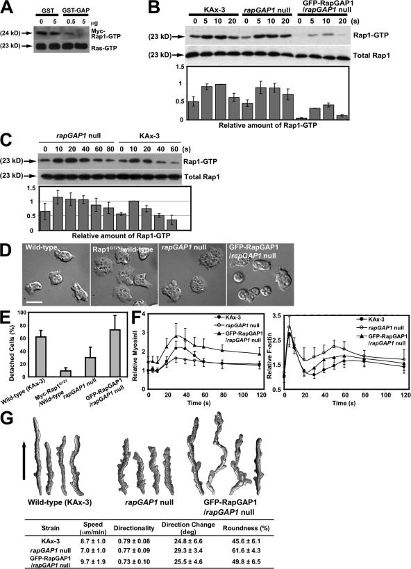
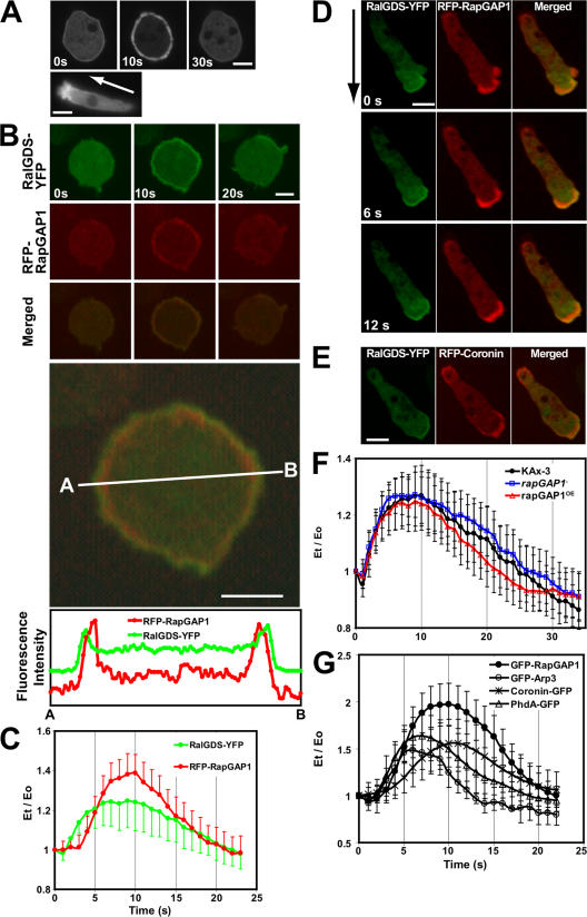
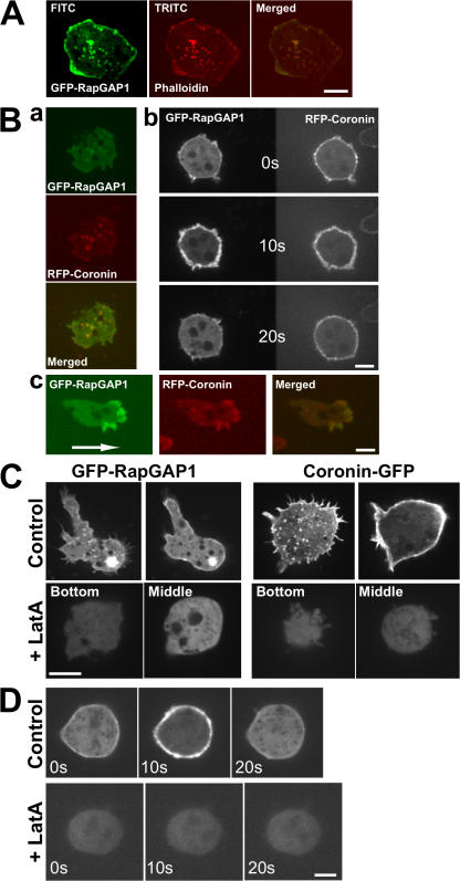
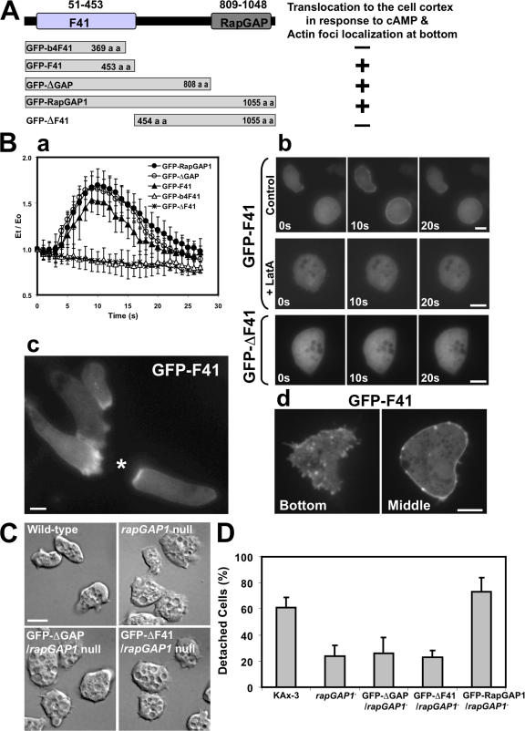
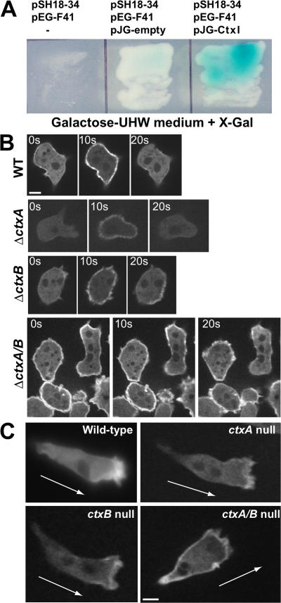
Spatial and temporal regulation of Rap1 is required for proper myosin assembly and cell adhesion during cell migration in Dictyostelium discoideum. Here, we identify a Rap1 guanosine triphosphatase-activating protein (GAP; RapGAP1) that helps mediate cell adhesion by negatively regulating Rap1 at the leading edge. Defects in spatial regulation of the cell attachment at the leading edge in rapGAP1- (null) cells or cells overexpressing RapGAP1 (RapGAP1(OE)) lead to defective chemotaxis. rapGAP1- cells have extended chemoattractant-mediated Rap1 activation kinetics and decreased MyoII assembly, whereas RapGAP1(OE) cells show reciprocal phenotypes. We see that RapGAP1 translocates to the cell cortex in response to chemoattractant stimulation and localizes to the leading edge of chemotaxing cells via an F-actin-dependent pathway. RapGAP1 localization is negatively regulated by Ctx, an F-actin bundling protein that functions during cytokinesis. Loss of Ctx leads to constitutive and uniform RapGAP1 cortical localization. We suggest that RapGAP1 functions in the spatial and temporal regulation of attachment sites through MyoII assembly via regulation of Rap1-guanosine triphosphate.


Figures

Figure 1.
Figure 1.
RapGAP1 negatively regulates cell adhesion and cell spreading. (A) Rap1-GTP stability assay using the GST fusion GAP domain of RapGAP1. (B) Rap1 activation kinetics in response to chemoattractant stimulation. (C) Extended time points from B (Materials and methods). (D) Spreading morphology of vegetative cells. Bar, 10 μm. (E) Cell–substratum adhesion assays. Adhesion was measured by the ratio of detached cells to the total number of cells. (F) Kinetics of F-actin polymerization and MyoII assembly in the Triton X-100–insoluble cytoskeletal fraction of the cells in response to chemoattractant stimulation. (G) Analysis of chemotaxis using DIAS software. Representative stacked images are shown. Superimposed images were taken every 1 min. The arrow indicates the direction of movement. Data represents mean ± SD from at least three experiments.
Figure 2.
Figure 2.
Localization of RapGAP1. (A) Translocation of GFP-RapGAP1 in wild-type cells to the cell cortex in response to uniform chemoattractant stimulation and localization of GFP-RapGAP1 during chemotaxis (bottom). The arrow indicates the direction of movement. (B–D) Dual-view analyses of the cells expressing both RalGDS-YFP and RFP-RapGAP1. (B) Translocations of the two proteins to the cell cortex by chemoattractant stimulation were contemporaneously imaged using a dual-view splitter. The merged image at 10 s after cAMP stimulation was enlarged in the bottom picture and the fluorescence intensities were measured along a line through the central portion of the cell. (C) Translocation kinetics of RalGDS-YFP and RFP-RapGAP1 to the cell cortex. (D and E) Spatial localizations of RalGDS-YFP and RFP-RapGAP1 (D) or RFP-coronin (E) in chemotaxing cells. The arrow indicates the direction of movement. (F) The translocation kinetics of RalGDS-YFP to the cell cortex in response to chemoattractant stimulation. (G) Translocation kinetics of GFP-RapGAP1, PhdA-GFP, GFP-Arp3, and coronin-GFP from time-lapse recordings, which were quantitated as described previously (Jeon et al., 2007). The graphs are the means of several cells from videos from at least three separate experiments. The graphs in C, F, and G represent means of >10 cells from at least three separate experiments done on different days. Error bars represent SD. Bars, 5 μm.
Figure 3.
Figure 3.
Colocalization of RapGAP1 with F-actin. (A) F-actin staining of the bottom sections of fixed vegetative cells expressing GFP-RapGAP1 using confocal microscopy. (B) Dual views of cells coexpressing GFP-RapGAP1 and RFP-coronin. (a) Bottom sections of the cells. (b) Translocation of GFP-RapGAP1 and RFP-coronin in response to uniform cAMP stimulation. (c) Localization of the two proteins during chemotaxis. An arrow indicates the direction of movement during chemotaxis. (C) Effect of LatA on localization of GFP-RapGAP1 and coronin-GFP. (D) Translocation of GFP-RapGAP1 to the cell cortex in the presence of LatA. Bars, 5 μm.
Figure 4.
Figure 4.
Cortical localization of RapGAP1 through the F41 domain. (A) Schematic diagram of truncated RapGAP1 proteins. (B) Translocation and localization of the truncated RapGAP1 proteins. (a) Translocation kinetics of the truncated GFP-fusion RapGAP1 proteins to the cell cortex in response to cAMP stimulation. (b) Translocation of GFP-F41 to the cell cortex upon stimulation without or with treatment of the cells with LatA. (c) Localization of GFP-F41 in chemotaxing cells. *, position of micropipette containing cAMP. (d) Foci localization of GFP-F41 at the bottom of cells. Cell morphology (C) and adhesion (D) of rapGAP1 cells expressing GFP-ΔF41 or GFP-ΔGAP. Experiments were performed at least three times. Error bars represent SD. Bars, 5 μm.
Figure 5.
Figure 5.
Localization of GFP-RapGAP1 in ctx-null cells. (A) Yeast two-hybrid analysis of the interaction of the F41 domain with CtxI. (B) Translocation of GFP-RapGAP1 in ctxA /B cells to the cell cortex in response to cAMP stimulation. (C) Localization of GFP-RapGAP1 in chemotaxing ctxA /B cells. The arrow indicates the direction of movement. Bars, 5 μm.

Similar articles

Cited by

References

    1. Bos, J.L. 2005. Linking Rap to cell adhesion. Curr. Opin. Cell Biol. 17:123–128. - PubMed
    1. Brinkmann, T., O. Daumke, U. Herbrand, D. Kuhlmann, P. Stege, M.R. Ahmadian, and A. Wittinghofer. 2002. Rap-specific GTPase activating protein follows an alternative mechanism. J. Biol. Chem. 277:12525–12531. - PubMed
    1. Chisholm, R.L., and R.A. Firtel. 2004. Insights into morphogenesis from a simple developmental system. Nat. Rev. Mol. Cell Biol. 5:531–541. - PubMed
    1. Chung, C.Y., and R.A. Firtel. 1999. PAKa, a putative PAK family member, is required for cytokinesis and the regulation of the cytoskeleton in Dictyostelium discoideum cells during chemotaxis. J. Cell Biol. 147:559–576. - PMC - PubMed
    1. Cornillon, S., L. Gebbie, M. Benghezal, P. Nair, S. Keller, B. Wehrle-Haller, S.J. Charette, F. Bruckert, F. Letourneur, and P. Cosson. 2006. An adhesion molecule in free-living Dictyostelium amoebae with integrin beta features. EMBO Rep. 7:617–621. - PMC - PubMed

Publication types

MeSH terms