. 2008 Feb;7(2):212-22.
doi: 10.1128/EC.00361-07. Epub 2007 Nov 26.

Target of rapamycin and LST8 proteins associate with membranes from the endoplasmic reticulum in the unicellular green alga Chlamydomonas reinhardtii

Affiliations

Target of rapamycin and LST8 proteins associate with membranes from the endoplasmic reticulum in the unicellular green alga Chlamydomonas reinhardtii

Sandra Díaz-Troya et al. Eukaryot Cell. 2008 Feb.

Abstract

The highly conserved target of rapamycin (TOR) kinase is a central controller of cell growth in all eukaryotes. TOR exists in two functionally and structurally distinct complexes, termed TOR complex 1 (TORC1) and TORC2. LST8 is a TOR-interacting protein that is present in both TORC1 and TORC2. Here we report the identification and characterization of TOR and LST8 in large protein complexes in the model photosynthetic green alga Chlamydomonas reinhardtii. We demonstrate that Chlamydomonas LST8 is part of a rapamycin-sensitive TOR complex in this green alga. Biochemical fractionation and indirect immunofluorescence microscopy studies indicate that TOR and LST8 exist in high-molecular-mass complexes that associate with microsomal membranes and are particularly abundant in the peri-basal body region in Chlamydomonas cells. A Saccharomyces cerevisiae complementation assay demonstrates that Chlamydomonas LST8 is able to functionally and structurally replace endogenous yeast LST8 and allows us to propose that binding of LST8 to TOR is essential for cell growth.


Figures

FIG. 1.
FIG. 1.
(A) Amino acid sequence and alignment of the seven WD-40 repeats in CrLST8. Residues that meet the criteria for a consensus WD repeat (48) are highlighted in gray. Each WD repeat consists of four β-strands, indicated by arrows A, B, C, and D. Critical residues for CrLST8 function identified in our screen are typed in red. Mutations identified in yeast and mammalian LST8 are typed in green and yellow, respectively. Mutations that were originally isolated in yeast LST8 (33, 42) and subsequently reproduced in mLST8 (27) are typed in blue. (B) Modeling analysis of CrLST8. The model was created by Swiss-Model (17), based on the homology of CrLST8 and the C-terminal WD-40 domain of TUP1 and LIS1/ALFA2. The image was prepared by using MOLMOL software. Red dots indicate the location of D106G, T228A, and S230N mutations. (C) Neighbor-joining tree of LST8s. The bootstrap values represent 1,000 replications. Genes used in the phylogenetic analysis of LST8s are as follows: C. reinhardtii, gwH.54.116.1 (ChlamyDB locus name); A. thaliana LST8.1, At3g18140 (TIGR locus name); A. thaliana LST8.2, At2g22040 (TIGR locus name); O. lucimarinus, eugene.0600010187 (JGI locus name); O. sativa, Os03g47780 (TIGR locus name); S. cerevisiae, LST8; S. pombe, Wat1/Pop3; D. melanogaster, CG3004 (FlyBase gene name); and H. sapiens, mLST8.
FIG. 2.
FIG. 2.
Localization of CrTOR and CrLST8 in large, membrane-associated complexes. (A) Whole-cell extracts were made from wild-type cells and probed with CrTOR and CrLST8 antibodies or preimmune antisera (pre). Arrows indicate single bands at the predicted molecular masses of ∼280 and ∼35 kDa recognized by CrTOR and CrLST8 antibodies, respectively. (B) Gel filtration elution profiles of CrTOR and CrLST8. Whole-cell lysates prepared from wild-type cells were loaded onto a Superose 6 sizing column. Fractions (0.5 ml) were collected and processed for immunodetection of CrTOR and CrLST8. The elution patterns of known molecular mass standards are indicated. (C) Fraction of CrTOR present in an SE versus total CrTOR in the cell. SE was prepared as described in Materials and Methods by freezing and thawing cells. Intact cells were removed from the cellular debris pellet by centrifugation at 400 × g for 1 min. The remaining insoluble membranous fraction was washed three times with PBS buffer and then treated or not (control) with salt and/or a nonionic detergent for 30 min on ice. The amount of CrTOR present in the SE, in the solubilized material, and in the remaining pellet (P) from the untreated control sample was monitored by Western blotting. (D) A portion of CrTOR and CrLST8 associate with microsomes. Biochemical fractionation of Chlamydomonas cell extracts was performed as described in Materials and Methods. Total SE was subjected to ultracentrifugation to separate soluble proteins that remain in the supernatant (S) from proteins that associate with the pelleted, membranous fraction (P). SEs were treated or not (control) with agents that disrupt membranes and membrane-protein interactions for 1 h at 4°C prior to ultracentrifugation. The presence of CrTOR, CrLST8, CrFKBP12, COXIIb, FOX1, the ER marker protein BiP, a vacuolar membrane ATPase (vATPase), and the chloroplast membrane protein D1 in these fractions was examined by Western blotting. D1 and COXIIb localization were also analyzed in the insoluble membrane fraction (M) that results after cell lysis. The asterisk indicates a presumed degradation product of CrLST8. (E) Sucrose density gradient profiles of CrTOR, CrLST8, BiP, vATPase, and FOX1. The microsomal fraction was fractionated on a 12-ml, 16 to 55% sucrose gradient. Fractions (1 ml) were collected from the top of the gradient and processed for Western blot analysis.
FIG. 3.
FIG. 3.
CrLST8 interacts with CrTOR through its kinase domain. (A) A 25-μg portion of purified GST-CrFKBP12 was bound to rapamycin and incubated with 1 mg of Chlamydomonas total extracts in the presence or absence of the reversible cross-linker DTSSP (for details, see Materials and Methods). Fusion protein complexes were immobilized on glutathione-Sepharose 4B beads and resolved by SDS-PAGE. CrTOR and CrLST8 were detected by Western blotting. (B) Pull-down assays were performed with the kinase domain of CrTOR fused to MBP (MBP-CrTORkinase) or MBP alone and total SE from Chlamydomonas cells. About 10 μg of MBP or MBP fusion protein were incubated with 4 mg of Chlamydomonas total extract. Endogenous CrLST8 bound to MBP-kin was detected by Western blotting with CrLST8 antibody.
FIG. 4.
FIG. 4.
Immunofluorescence localization of CrTOR, CrLST8, and BiP proteins in Chlamydomonas cells. (A) Exponentially growing wild-type cells were collected and processed for IF microscopy analysis as described in Materials and Methods. The antigens of interest are shown in green (FITC), whereas DNA staining is shown in blue (DAPI). Merge images show a merge of the green and blue channels. (B) Double staining of α-tubulin and CrTOR (top) or CrLST8 (down). CrTOR and CrLST8 are shown in green (FITC), α-tubulin in red (tubulin), and DNA in blue (DAPI). Merge images show a merge of the green, red, and blue channels.
FIG. 5.
FIG. 5.
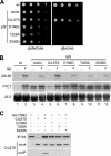
CrLST8 functionally and structurally substitutes yeast LST8. (A) CrLST8 complements a yeast lst8 mutant. Wild-type TB50a cells were transformed with empty vector (wt). Cells expressing LST8 from the glucose-repressible and galactose-inducible GAL1 promoter were transformed empty vector (vector) or with plasmids expressing wild-type CrLST8 or D106G, T228A, or S230N CrLST8 mutants. Cells were grown in glucose-containing, liquid medium for 15 h, normalized, subjected to 10-fold serial dilutions, and spotted onto SGal (galactose) or SD (glucose) plates. (B) CrLST8 is able to regulate the TOR-controlled transcription factors GLN3 and RTG1/3. Wild-type cells and GAL1-driven lst8 cells expressing wild-type CrLST8 or the different CrLST8 mutants were grown for 15 h in SD medium (for the analysis of GLN3 targets) or in SD medium supplemented with 3% l-glutamine (for the analysis of RTG1/3 targets). Cells were treated with drug vehicle (90% ethanol-10% Tween 20) or rapamycin for 30 min. Total RNA was probed with 32P-labeled DNA probes specific for the GLN3 target DAL80 or the RTG1/3 target PYC1. 25S rRNA was used as a loading control. (C) Wild-type CrLST8 coimmunoprecipitates with yeast TOR2. A yeast strain expressing HA-tagged TOR2 and GAL1-driven LST8 was transformed with plasmids harboring wild-type or mutant CrLST8s. Cells were depleted of endogenous LST8 by incubation for 15 h in SD medium. Lysates were prepared and subjected to immunoprecipitation with anti-HA. Immunoprecipitates were probed with anti-CrLST8 to detect immunoprecipitated CrLST8 (lower panel).

Similar articles

Cited by

References

    1. Anderson, G. H., and M. R. Hanson. 2005. The Arabidopsis Mei2 homologue AML1 binds AtRaptor1B, the plant homologue of a major regulator of eukaryotic cell growth. BMC Plant Biol. 52-9. - PMC - PubMed
    1. Anderson, G. H., B. Veit, and M. R. Hanson. 2005. The Arabidopsis AtRaptor genes are essential for post-embryonic plant growth. BMC Biol. 312-22. - PMC - PubMed
    1. Aronova, S., K. Wedaman, S. Anderson, J. Yates III, and T. Powers. 2007. Probing the membrane environment of the TOR kinases reveals functional interactions between TORC1, actin, and membrane trafficking in Saccharomyces cerevisiae. Mol. Biol. Cell 182779-2794. - PMC - PubMed
    1. Burnett, P. E., R. K. Barrow, N. A. Cohen, S. H. Snyder, and D. M. Sabatini. 1998. RAFT1 phosphorylation of the translational regulators p70 S6 kinase and 4E-BP1. Proc. Natl. Acad. Sci. USA 951432-1437. - PMC - PubMed
    1. Cardenas, M. E., and J. Heitman. 1995. FKBP12-rapamycin target TOR2 is a vacuolar protein with an associated phosphatidylinositol-4 kinase activity. EMBO J. 145892-5907. - PMC - PubMed

Publication types

MeSH terms

LinkOut - more resources