. 2007 Dec;134(24):4449-58.
doi: 10.1242/dev.012187.

Obligatory participation of macrophages in an angiopoietin 2-mediated cell death switch

Affiliations

Obligatory participation of macrophages in an angiopoietin 2-mediated cell death switch

Sujata Rao et al. Development. 2007 Dec.

Abstract

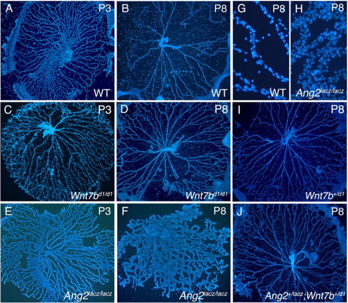
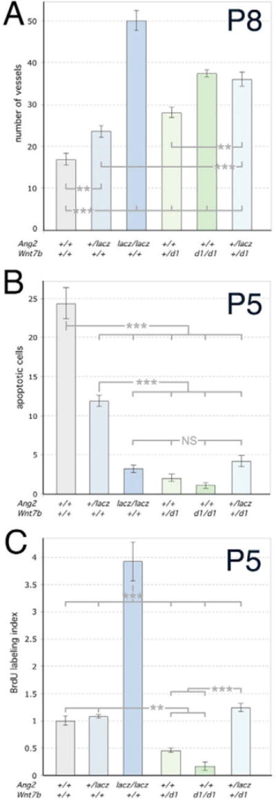
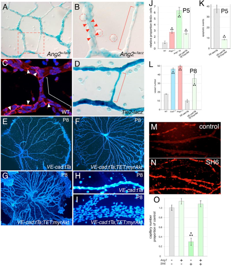
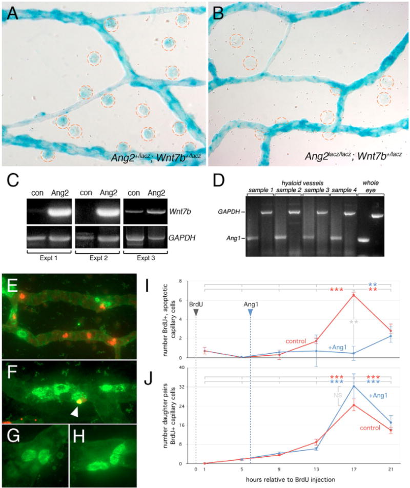
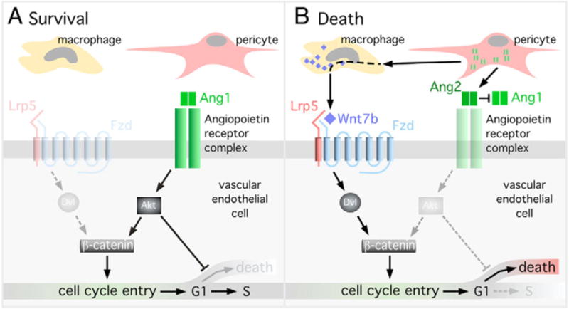
Macrophages have a critical function in the recognition and engulfment of dead cells. In some settings, macrophages also actively signal programmed cell death. Here we show that during developmentally scheduled vascular regression, resident macrophages are an obligatory participant in a signaling switch that favors death over survival. This switch occurs when the signaling ligand angiopoietin 2 has the dual effect of suppressing survival signaling in vascular endothelial cells (VECs) and stimulating Wnt ligand production by macrophages. In response to the Wnt ligand, VECs enter the cell cycle and in the absence of survival signals, die from G1 phase of the cell cycle. We propose that this mechanism represents an adaptation to ensure that the macrophage and its disposal capability are on hand when cell death occurs.


Figures

Fig. 1
Fig. 1. Genetic Interaction between the Wnt and the angiopoietin pathways
(A–J) Hyaloid vessels from WT (A,B,G), Wnt7bd1/d1 (C,D), Ang2lacZ/lacZ (E,F,H), Wnt7b+/d1 (I) and Ang2+/lacZ; Wnt7b+/d1 (J) animals of the indicated ages stained with Hoechst 33258. (H) Hypercellularity of Ang2lacZ/lacZ hyaloid capillaries compared with WT (G) at P8. Magnifications: 50 × in A–F,I,J; 400 × in G,H.
Fig. 2
Fig. 2. The Wnt and angiopoietin pathways are finely balanced in regulating cell death, proliferation and hyaloid regression
(A–C) Numbers of hyaloid vessels at P8 (A), apoptotic cells at P5 (B) and BrdU-labeled cells at P5 (C) in animals of the indicated genotypes. Significance was assessed using one-way ANOVA. All genotypes were compared to WT and were significantly different as indicated by the asterisks. Brackets indicate the groups that were compared using oneway ANOVA. Significance levels: **P ≤ s0.01, > 0.001; ***P ≤ s0.001, > 0.
Fig. 3
Fig. 3. Angiopoietin pathway responses downstream of Akt regulate hyaloid regression
(A,B) X-Gal staining of P5 hyaloid vessels from Ang2+/lacZ mice. Dashed red box in A indicates region shown at higher magnification in B. Red arrowheads indicate X-Gal-labeled cell processes, the red bracket indicates a small capillary with limited X-Gal staining. Macrophages are indicated by the dashed red circles. (C) Pericytes from P5 hyaloid vessels of wild-type mice labeled with desmin (red). Nuclei are stained with Hoechst 33258 (blue). White arrowheads indicate cell processes of the pericytes, the white bracket indicates areas with no desmin staining. (D) X-Gal staining of P5 hyaloid vessels from a Tie2-lacZ mouse in which lacZ is expressed in vascular endothelial cells (VECs). The red bracket indicates a small capillary with uniform X-Gal staining. (E–I) Hoechst 33258-labeled hyaloid vessels from control (E,H) and VE-cadherin:tTa; TET:myrAkt (F,G,I) transgenic animals. (J) Proportion of BrdU-positive cells in P5 hyaloid vessels of WT, Ang2lacZ/lacZ, WT injected with Ang1 and VE-cadherin:tTa;TET:myrAkt mice. (K) Number of apoptotic events at P5 in the hyaloid vessels from mice of the indicated genotypes. (L) Hyaloid vessel number at P8 in mice of the indicated genotypes. (M,N) TUNEL labeling of hyaloid vessel preparations with no treatment (M) or after intravitreal injection of SH6 (N). (O) Quantification of healthy hyaloid vessel number as a proportion of wild type after injection of the indicated reagents at P4 and assessment at P5. Magnifications: 50 × (E–G), 200 × (A), 400 × (M,N), 630 × (B,C,D,H,I). Significance levels: **P ≤ s0.01, > 0.001; ***P ≤ s0.001, > 0.
Fig. 4
Fig. 4. The Tie2 and canonical Wnt pathways are integrated through Akt
(A–E) Immunofluorescence detection of β-catenin in primary MVECs either untreated (A) or treated with Wnt3a (B), Ang1 (C), Ang1 + wortmannin (D) or Ang1 + SH6 (E). Upper and lower parts of each panel show β-catenin labeling alone and merged nuclear and β-catenin labeling, respectively. (F) Proportion of MVECS with nuclear β-catenin detectable by immunofluorescence under the conditions indicated. For this quantification, the significance levels are: **P < 0.005, ****P < 0.00005.
Fig. 5
Fig. 5. The angiopoietin pathway activates Wnt target genes via Akt andβ-catenin
(A–L) Immunofluorescent staining for β-catenin (green and red with two rounds of labeling – see text) and nuclei (blue) in hyaloid preparations in mice of the indicated genotypes. (M,N) X-Gal staining of hyaloid vessels (red arrowheads) from TOPGAL; Lrp5+/lacZ animals that were (M) mock injected intra-vitreally or (N) injected with Ang1 intra-vitreally. (O,P) Quantification of X-Gal-stained cell numbers (as in M and N) from hyaloid vessels of the indicated genotypes injected with Ang1 (O) or Ang2 (P). Significance levels: **P ≤ s0.01, > 0.001.
Fig. 6
Fig. 6. Angiopoietin signaling regulates Wnt7b expression and cell cycle stage-dependent cell death
(A,B) X-Gal staining of hyaloid vessels from Ang2lacZ/+; Wnt7blacZ/+ (A) and Ang2lacZ/lacZ; Wnt7blacZ/+ (B) animals. Macrophages are circled in red. (C)Wnt7b and Gapdh RT-PCR of laser captured macrophages from hyaloid vessels of P3 wild-type animals, injected intra-vitreally with Ang2 (Ang2) or sham injected (con). Data from three independent experiments are presented. (D)Ang1 and Gapdh RT-PCR from wild-type P5 hyaloid vessel mRNA. Whole eye RNA was used as a control. (E–H) Combined BrdU (green) and TUNEL (red) labeling of hyaloid vessels showing BrdU-positive VECs that are apoptotic (F, indicated by white arrowhead) and pairs of BrdU-positive daughter nuclei (G,H). (I,J) BrdU labeled cell fate mapping: red lines show the number of BrdU-TUNEL double labeled nuclei (I) and BrdU-positive daughter pairs (J) in left eye hyaloid vessels assessed over a 21 hour time-course after BrdU incorporation. The blue lines in I and J show similar quantification in the right, contralateral eye after Ang1 injection at t = 6 hours. Significance levels: **P ≤ 0.01, > 0.001; ***P ≤ 0.001, > 0.
Fig. 7
Fig. 7. Model of the angiopoietin 2-dependent cell death switch
(A) Hyaloid VECs will survive when Ang1 activates Akt, suppresses cell death and promotes proliferation through β-catenin stabilization. (B) Hyaloid VECs will die when Ang2 promotes cell death via suppression of Akt and cell cycle entry by upregulating Wnt7b in macrophages. In the absence of survival signals the cycling hyaloid VECs will undergo apoptosis in the G1 phase of the cell cycle.

References

    1. Assoian RK. Anchorage-dependent cell cycle progression. J Cell Biol. 1997;136:1–4. - PMC - PubMed
    1. Camenisch G, Pisabarro MT, Sherman D, Kowalski J, Nagel M, Hass P, Xie MH, Gurney A, Bodary S, Liang XH. ANGPTL3 stimulates endothelial cell adhesion and migration via integrin alpha vbeta 3 and induces blood vessel formation in vivo. J Biol Chem. 2002;277:17281–17290. - PubMed
    1. Carlson TR, Feng Y, Maisonpierre PC, Mrksich M, Morla AO. Direct cell adhesion to the angiopoietins mediated by integrins. J Biol Chem. 2001;276:26516–26525. - PubMed
    1. Cascone I, Napione L, Maniero F, Serini G, Bussolino F. Stable interaction between alpha5beta1 integrin and Tie2 tyrosine kinase receptor regulates endothelial cell response to Ang-1. J Cell Biol. 2005;170:993–1004. - PMC - PubMed
    1. Castillo SS, Brognard J, Petukhov PA, Zhang C, Tsurutani J, Granville CA, Li M, Jung M, West KA, Gills JG. Preferential inhibition of Akt and killing of Akt-dependent cancer cells by rationally designed phosphatidylinositol ether lipid analogues. Cancer Res. 2004;64:2782–2792. - PubMed

Publication types