. 2007 Dec 4;104(49):19279-84.
doi: 10.1073/pnas.0709710104. Epub 2007 Nov 26.

Structure of the Nitrosomonas europaea Rh protein

Affiliations

Structure of the Nitrosomonas europaea Rh protein

Xin Li et al. Proc Natl Acad Sci U S A. .

Abstract

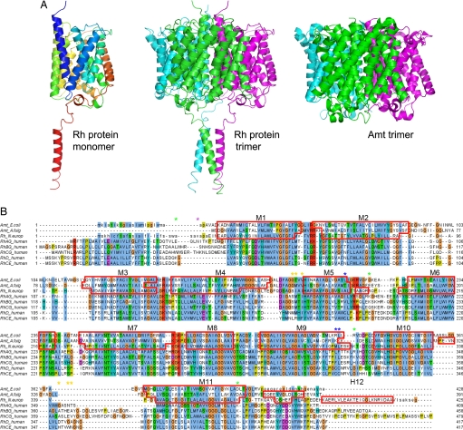
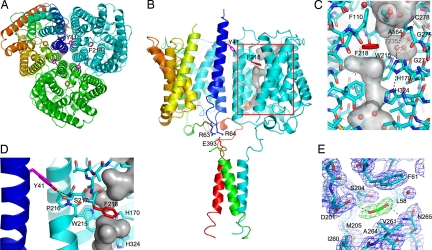
Amt/MEP/Rh proteins are a family of integral membrane proteins implicated in the transport of NH3, CH(2)NH2, and CO2. Whereas Amt/MEP proteins are agreed to transport ammonia (NH3/NH4+), the primary substrate for Rh proteins has been controversial. Initial studies suggested that Rh proteins also transport ammonia, but more recent evidence suggests that they transport CO2. Here we report the first structure of an Rh family member, the Rh protein from the chemolithoautotrophic ammonia-oxidizing bacterium Nitrosomonas europaea. This Rh protein exhibits a number of similarities to its Amt cousins, including a trimeric oligomeric state, a central pore with an unusual twin-His site in the middle, and a Phe residue that blocks the channel for small-molecule transport. However, there are some significant differences, the most notable being the presence of an additional cytoplasmic C-terminal alpha-helix, an increased number of internal proline residues along the transmembrane helices, and a specific set of residues that appear to link the C-terminal helix to Phe blockage. This latter linkage suggests a mechanism in which binding of a partner protein to the C terminus could regulate channel opening. Another difference is the absence of the extracellular pi-cation binding site conserved in Amt/Mep structures. Instead, CO2 pressurization experiments identify a CO2 binding site near the intracellular exit of the channel whose residues are highly conserved in all Rh proteins, except those belonging to the Rh30 subfamily. The implications of these findings on the functional role of the human Rh antigens are discussed.


Conflict of interest statement

The authors declare no conflict of interest.

Figures

Fig. 1.
Fig. 1.
Structure of the N. europaea Rh protein. (A Left) Ribbon diagram of N. europaea Rh protein subunit colored in rainbow from blue to red. (A Center) Ribbon diagram of N. europaea Rh protein trimer colored by subunit. (A Right) Ribbon diagram of E. coli AmtB colored by subunit. Note the additional C-terminal helix on the Rh protein. The diagram is oriented such that the periplasmic surface is on the top and the cytoplasmic face is on the bottom. (B) Sequence alignment of the N. europaea Rh protein to the E. coli AmtB and A. fulgidus Amt1 based on their respective structures and to the five human Rh proteins based on the program ClustalW (34). The residues not observed in the Rh protein structure are shown in lowercase. Segments assigned to α-helices are framed with red lines. Asterisks mark topologically relevant residues: glycosylation and protease sensitive sites on RhAG are indicated with purple and green asterisks, respectively; highly antigenic and potentially palmitoylated residues of RhD are labeled with orange and blue asterisks.
Fig. 2.
Fig. 2.
Functionally relevant sites within the N. europaea Rh protein. (A) Periplasmic view of the N. europaea Rh protein along the three-fold axis. Two key residues implicated in channel opening are colored in violet (Tyr-41) and red (Phe-218). Subunits are colored in cyan, green, and rainbow [from blue (N terminus) to red (C terminus)]. (B) Side view of the N. europaea Rh protein emphasizing the key residues implicated in partner protein-mediated channel gating. In the cyan subunit the surface of the channel is shown together with the location of the putative CO2 binding site. Tyr-41 on helix M1 from one subunit (in rainbow) is proposed to regulate the extent of kinking of transmembrane helix M6 on the adjacent subunit (in cyan). The position of Tyr-41 in turn is linked to the position of helix M1. Partner protein binding to the C-terminal α-helix is proposed to alter the position of the M1 helix via salt bridge interactions between Glu-393 on the C-terminal helix and Arg-63 and Arg-64 on helix M1. (C) Key channel residues on the periplasmic side of the channel. The protein is depicted in stick form with carbon atoms colored in cyan and the remaining atoms in CPK. Waters and the putative ethylene glycol are shown in ball-and-stick form. Potential cavities are highlighted with a transparent surface colored in gray. For the unique hydrophilic pocket adjacent to Phe-218, the hydrogen-bonding interactions of its three waters are depicted as black dotted lines. (D) Interactions between Tyr-41 and the M6 helix of the adjacent subunit. Potential hydrogen bonds between Tyr-41 and Ser-217 are shown as black dotted lines. (E) Weighted density maps surrounding the CO2 binding site, 2mFODFC (in black, contoured at 1.0σ) and mFODFC (in green, contoured at 3.0σ, and in red, contoured at −3.0σ) with terms as determined by REFMAC5 (36).

Similar articles

Cited by

References

    1. Avent ND, Reid ME. Blood. 2000;95:375–387. - PubMed
    1. Van Kim CL, Colin Y, Cartron JP. Blood Rev. 2006;20:93–110. - PubMed
    1. Ludewig U. Transfus Clin Biol. 2006;13:111–116. - PubMed
    1. Kustu S, Inwood W. Transfus Clin Biol. 2006;13:103–110. - PubMed
    1. Marini AM, Urrestarazu A, Beauwens R, Andre B. Trends Biochem Sci. 1997;22:460–461. - PubMed

Publication types

LinkOut - more resources