. 2007 Dec 4;104(49):19512-7.
doi: 10.1073/pnas.0709443104. Epub 2007 Nov 26.

Small molecule obatoclax (GX15-070) antagonizes MCL-1 and overcomes MCL-1-mediated resistance to apoptosis

Affiliations

Small molecule obatoclax (GX15-070) antagonizes MCL-1 and overcomes MCL-1-mediated resistance to apoptosis

Mai Nguyen et al. Proc Natl Acad Sci U S A. .

Abstract

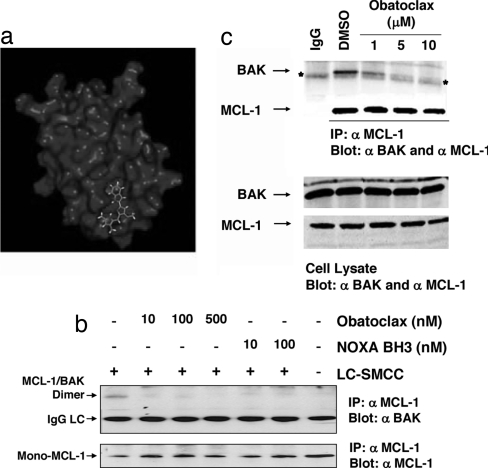
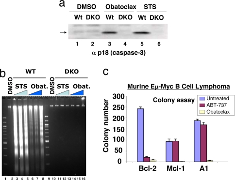
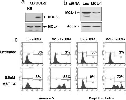
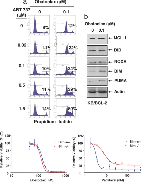
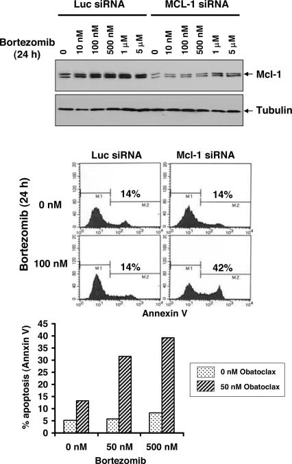
Elevated expression of members of the BCL-2 pro-survival family of proteins can confer resistance to apoptosis in cancer cells. Small molecule obatoclax (GX15-070), which is predicted to occupy a hydrophobic pocket within the BH3 binding groove of BCL-2, antagonizes these members and induces apoptosis, dependent on BAX and BAK. Reconstitution in yeast confirmed that obatoclax acts on the pathway and overcomes BCL-2-, BCL-XL-, BCL-w-, and MCL-1-mediated resistance to BAX or BAK. The compound potently interfered with the direct interaction between MCL-1 and BAK in intact mitochondrial outer membrane and inhibited the association between MCL-1 and BAK in intact cells. MCL-1 has been shown to confer resistance to the BCL-2/BCL-XL/BCL-w-selective antagonist ABT-737 and to the proteasome inhibitor bortezomib. In both cases, this resistance was overcome by obatoclax. These findings support a rational clinical development opportunity for the compound in cancer indications or treatments where MCL-1 contributes to resistance to cell killing.


Conflict of interest statement

Conflict of interest statement: R.C.M., A.R., M.W., L.S., S.R.M.M., D.G., J.V., L.B., X.B., P.B., and G.C.S. are or were employees of Gemin X Biotechnologies, Inc.

Figures

Fig. 1.
Fig. 1.
Obatoclax interferes with MCL-1/BAK interactions. (a) Predicted orientation of obatoclax within the BH3 binding groove of BCL-2 (see also SI Fig. 7 and SI Materials and Methods). (b) Disruption of constitutive MCL-1/BAK interactions in isolated mitochondria. Mitochondria were isolated from SK-Mel5 cells and incubated with the indicated concentrations of obatoclax or NOXA BH3 peptide for 30 min at 37°C and then subjected to chemical cross-linking with 0.1 mM LC-SMCC (+) or vehicle (DMSO) (−). After blocking excess LC-SMCC, the mitochondria were lysed with detergent and subjected to immunoprecipitation with anti MCL-1 antibody, and the precipitates were probed by immunoblot with anti-MCL-1 and anti BAK antibodies. (c) Obatoclax antagonizes MCL-1/BAK association in cells. SK-Mel5 cells were incubated with DMSO or obatoclax in DMSO for 5 h, after which time total cell lysate was subjected to immunoprecipitation with anti MCL-1 antibody. Immunoprecipitates and cell lysates were separated by SDS/PAGE and analyzed by Western blot with anti-MCL-1 and anti-BAK antibodies.
Fig. 2.
Fig. 2.
Induction of apoptosis and inhibition of colony formation by obatoclax. (a) Deletion of Bax and Bak causes resistance to obatoclax. Baby kidney epithelial cell lines derived from the WT and Bax,Bak double knockout (DKO) mouse were treated with vehicle (DMSO) or vehicle containing 2.0 μM obatoclax or 0.5 μM staurosporin (STS), and 20 h later the processed large subunit of caspase-3 (arrow) was detected by SDS/PAGE and immunoblot analysis. (b) As in a, except that oligonucleosomal fragments were detected by agarose gel electrophoresis and staining with ethidium bromide after treatments with 0.05 μM (lanes 3 and 10), 0.2 μM (lanes 4 and 11) and 1 μM (lanes 5 and 12) STS for 16 h; obatoclax treatment was 0.5 μM (lanes 6 and 14), 1 μM (lanes 7 and 15) and 2 μM (lanes 8 and 16) for 24 h. (c) Sensitivity of murine Eμ Myc B cell lymphoma cell lines overexpressing Bcl-2, Mcl-1, or A1 to obatoclax. Cells were exposed to 1 μM obatoclax or ABT737 for 24 h, the media (containing compound) diluted 30-fold, and colony formation on soft agar recorded after 7 days, as described in ref. .
Fig. 3.
Fig. 3.
BCL-2-overexpressing KB cells whose endogenous MCL-1 confers resistance to ABT-737. (a) Stable overexpression of BCL-2 in KB cells. (b) Reduction of MCL-1 expression in KB/BCL-2 cells by MCL-1 siRNA. (c) MCL-1 knockdown confers sensitivity to ABT-737 in KB/BCL-2 cells. KB/BCL-2 cells were transfected with human MCL-1 siRNA or luciferase (Luc) siRNA and plated into media containing 0.5 μM ABT-737. After 72, the percentage of cells incorporating annexin V FITC and propidium iodide was determined by flow cytometry.
Fig. 4.
Fig. 4.
Obatoclax overcomes the resistance of KB/BCL-2 cells to ABT-737. (a) KB/BCL-2 cells were incubated with the indicated concentrations of ABT-737 in the absence or presence of 0.1 μM obatoclax. The percentage of cells that took up propidium iodide was determined by flow cytometry. (b) Influence of obatoclax on expression of MCL-1 and BH3-only antagonists of MCL-1 in KB/BCL-2 cells. After treatment with or without 0.1 μM obatoclax for 72 h, 25 μg of total cell lysate was analyzed by SDS/PAGE and immunoblot for the indicated proteins. (c) Bim deletion does not interfere with obatoclax cytotoxicity. Bim−/− or Bim+/+ transformed baby mouse kidney cells were treated with obatoclax or paclitaxel at the indicated concentrations for 72 h and cell viability determined. Standard error is shown (bars).
Fig. 5.
Fig. 5.
Obatoclax overcomes MCL-1-mediated resistance to bortezomib in melanoma cells. (Top) Mouse melanoma B16-F1 cells were transfected with luciferase siRNA or mouse Mcl-1 siRNA for 24 h followed by a 24-h treatment with bortezomib at the indicated concentrations. Total cell lysates were analyzed by immunoblot for Mcl-1 expression. (Middle) As in Top, except that the percentage of cells that incorporated annexin V was determined by flow cytometry. (Bottom) B16-F1 cells were incubated with bortezomib alone or in combination with 50 nM obatoclax for 24 h and apoptosis (annexin V staining) determined as in Middle.

Similar articles

Cited by

References

    1. Cory S, Vaux DL, Strasser A, Harris AW, Adams JM. Cancer Res. 1999;59(Suppl 7):1685s–1692s. - PubMed
    1. Korsmeyer SJ. Cancer Res. 1999;59(Suppl 7):1693s–1700s. - PubMed
    1. Danial NN, Korsmeyer SJ. Cell. 2004;116:205–219. - PubMed
    1. Vaux DL, Cory S, Adams JM. Nature. 1988;335:440–442. - PubMed
    1. Reed JC, Cuddy M, Slabiak T, Croce CM, Nowell PC. Nature. 1988;336:259–261. - PubMed

Publication types

MeSH terms