. 2008 Apr;56(4):329-45.
doi: 10.1369/jhc.7A7321.2007. Epub 2007 Nov 26.

Immunolocalization of Periostin-like factor and Periostin during embryogenesis

Affiliations

Immunolocalization of Periostin-like factor and Periostin during embryogenesis

Shimei Zhu et al. J Histochem Cytochem. 2008 Apr.

Abstract

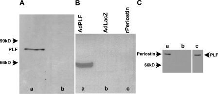
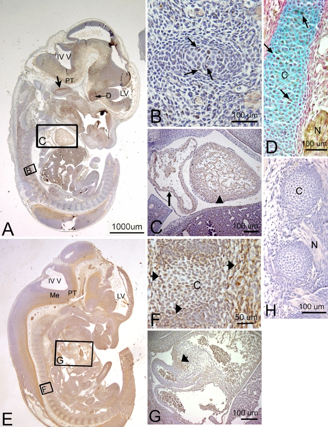
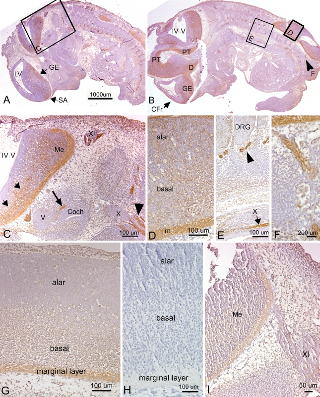
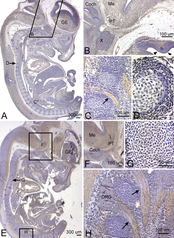
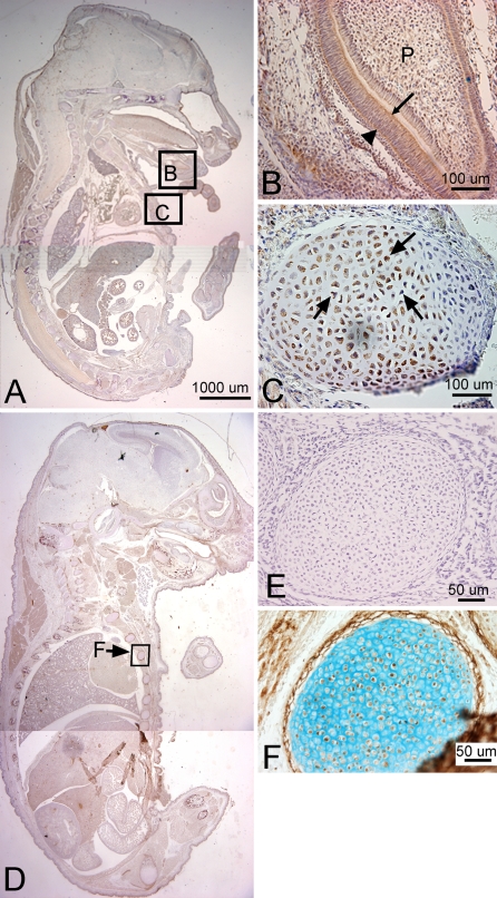
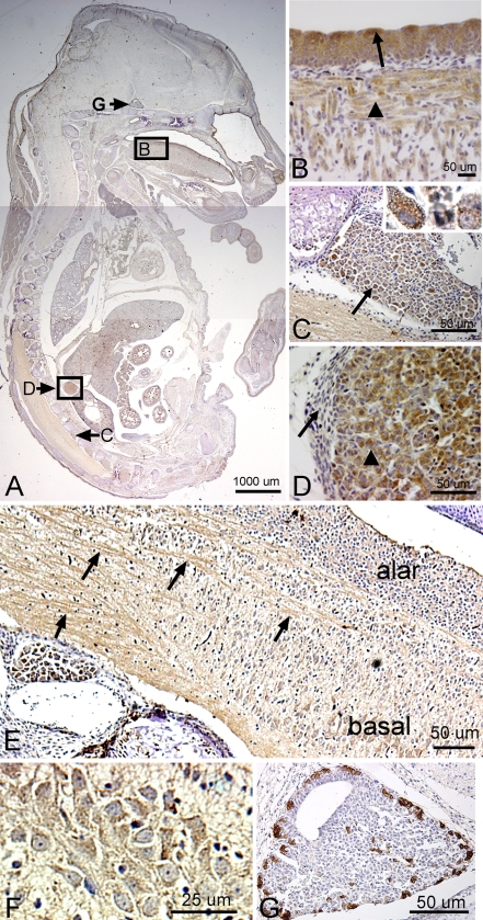
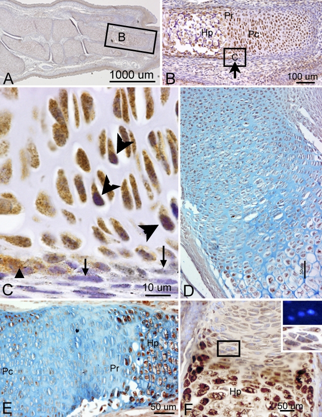
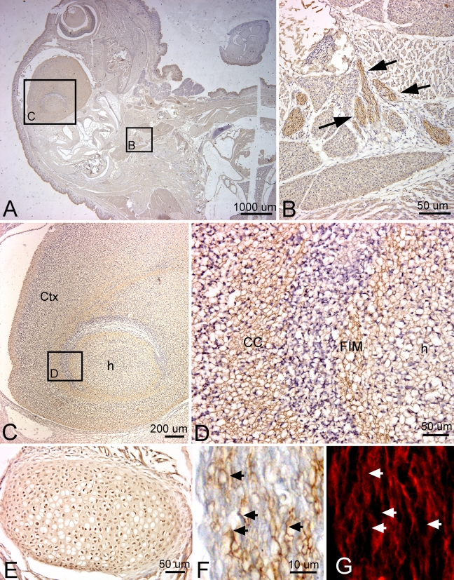
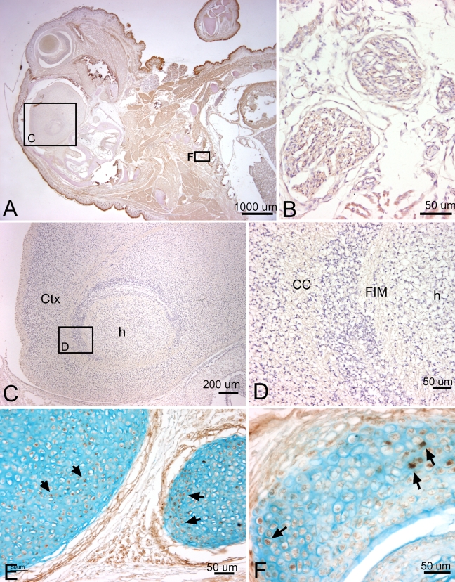
Periostin-like factor (PLF) and Periostin are alternatively spliced mRNAs. Our findings are the first to show similarities and differences between PLF and Periostin location using isoform-specific antibodies. The differences in when and where they are present during mouse embryogenesis suggest that they may have different functions. Using immunostaining techniques, we observed that PLF was highly expressed at 12.5 days postconception (dpc) in the intermediate and outer zones of most brain regions, spinal cord, cranial and spinal nerves, and chondrocytes in developing bone and in the heart wall. By 16.5 dpc, PLF was also present in ameloblasts and odontoblasts in developing teeth, and by 19.5 dpc, PLF was present at low levels only in vagal nerve bundles, discrete white matter bundles in the brain, and chondrocytes of developing ribs. Periostin, on the other hand, was absent at 12.5 dpc from dorsal spinal cord and from cranial and spinal nerves. By 16.5 dpc, Periostin was present in many spinal nerves, but absent thereafter, and at 19.5 dpc, Periostin was present in chondrocytes in developing bone but not in neural tissues. The different spatial and temporal location of PLF and Periostin in cartilage and bone cells suggests different roles for these proteins in endochondral bone formation. The early expression of PLF in brain differentiation zones and in developing axon bundles and nerves suggests that it may facilitate axon growth.


Figures

Figure 1
Figure 1
Specificity of Periostin-like factor (PLF) and Periostin-specific antibodies. (Aa) One hundred μg total protein from neonatal mouse heart was separated by SDS-PAGE followed by Western blot analysis. PLF antibody recognized one band at 90 kDa. (Ab) Competition of antibody with the antigenic peptide before Western blot analysis resulted in elimination of the 90-kDa band. (Ba) Total protein from COS cells in which PLF was overexpressed by adenovirus, (Bb) total protein from COS cells infected with control virus, and (Bc) recombinant Periostin (rPeriostin). Periostin, a PLF-related isoform, is not recognized by PLF antibody, confirming specificity of anti-PLF. (C) Western blot with Periostin antibody (Ca,Cb), and PLF antibody (Cc). (Ca) Total protein from the femur of 17.5-day-old mouse embryos probed with Periostin antibody. (Cb,Cc) Total protein from COS cells in which PLF was overexpressed by adenovirus: (Cb) probed with Periostin antibody and (Cc) probed with PLF antibody. PLF was not recognized by the Periostin antibody as shown in Cb.
Figure 2
Figure 2
Localization of PLF and Periostin proteins in developing cartilage and myocardium in a mouse embryo at 12.5 days postconception (dpc). Sectioned embryo was probed with PLF antibody (A–D) or Periostin antibody (E–G) and counterstained with hematoxylin (a nuclear stain, A–H), nuclear red (a nuclear stain, D), or Alcian blue (a cartilage stain, D). (A) Sagittal section of embryo at low magnification showing fourth (IV V) and lateral (LV) ventricles and positive PLF staining in the presumptive pyramidal track (PT). Large arrow indicates PLF stained cells lining cochlear lumen (brown); small arrow indicates PLF staining in optic stalk region of diencephalon (D). (B) Higher magnification of corresponding area in A showing cells weakly stained for PLF in developing vertebra (arrows). (C) Higher magnification of corresponding area in A showing PLF staining in the atrium (arrow) and ventricle (arrowhead) of a developing heart. (D) Arrows indicate PLF-stained cells (brown) in rib cartilage (C, blue) and in nearby peripheral nerve (N). (E) Sagittal section of embryo at low magnification showing Periostin staining in the medullary differentiating field (Me) and presumptive pyramidal track (PT). (F) Higher magnification of corresponding area in E showing mesenchymal cells that are stained weakly for Periostin in and around the developing vertebral cartilage (C, arrows). (G) Higher magnification of corresponding area in E showing Periostin staining in the atrium (arrow) of a developing heart. (H) Photo of negative antibody control slide (no primary antibody) showing an absence of PLF staining in cartilage (C) and nerve (N).
Figure 3
Figure 3
Localization of PLF (A–F) and Periostin (G,I) proteins in developing nervous system in mouse embryo at 12.5 days dpc, counterstained with hematoxylin. (A,B) Sagittal sections of embryo at low magnification showing PLF staining in the ganglionic eminence (GE) differentiating field, septal area (SA), diencephalon optic stalk area (D), frontal cortical differentiation field (CFr, arrow in B), and pyramidal track (PT). Arrowhead in B indicates PLF stained lumbosacral nerve also shown in F. IV V, fourth ventricle; LV, lateral ventricle. (C) Higher magnification of corresponding area in A showing axon bundles (small arrows) stained with PLF in presumptive pons and medulla (Me) and in neural processes of glossopharyngeal (XI) and vagus (X) cranial nerve ganglia (arrowhead) and in the cochlea (coch, large arrow), but not in the trigeminal ganglion (V). (D) Higher magnification of corresponding area in B showing PLF staining in spinal cord basal and alar plates and marginal zone (m). (E) Higher magnification of corresponding area in B showing intense PLF staining in segmental axon bundles (arrowhead) and vagal nerve (X, arrow) in the thorax. The dorsal root ganglion (DRG) is not stained at this stage. (F) Higher magnification of corresponding area in B showing PLF staining in peripheral lumbosacral nerve. (G) Higher magnification of Periostin staining in the spinal cord (low magnification shown in Figure 1E) showing Periostin staining in spinal cord basal plate and marginal layer, but not in the alar plate. (H) Photograph of negative antibody control slide (no primary antibody) showing an absence of Periostin staining. (I) A higher magnification of a hindbrain region showing Periostin staining in the marginal zone of the presumptive medulla (Me). Little to no staining is present in processes of the glossopharyngeal ganglion (XI).
Figure 4
Figure 4
Localization of PLF (A–D) and periostin (E–H) proteins in developing nervous system in mouse embryo at 13.5 days dpc, counterstained with hematoxylin. (A) Sagittal section of embryo at low magnification, with the ganglionic eminence (GE) indicated. PLF staining is less distinct, overall, in 13.5 dpc embryos than in 12.5 dpc embryos. (B) Higher magnification of corresponding area in A showing PLF staining in the hippocampus ventricular zone (arrowheads) and differentiating zone (h), processes from the vagus ganglion (X), and within the cochlea (Coch). The presumptive pyramidal tract (PT) and medulla (Me) are faintly stained. (C) Higher magnification from a section adjacent to section shown in A (C*), but a similar region, showing intense PLF staining in lumbosacral nerves (arrow) passing through developing vertebrae. (D) Higher magnification of area indicated by arrow in A showing only faint PLF staining in a developing vertebral body located at the mid-thoracic level. (E) Sagittal section of embryo at low magnification, showing Periostin staining in several nervous system regions. (F) Higher magnification of corresponding area in E showing Periostin staining the presumptive pyramidal tract (PT) and medulla (Me) but not the cochlea (Coch). (G) Higher magnification of area indicated by arrow in E showing Periostin staining cells in a developing vertebral body. (H) Higher magnification of corresponding area indicated in E showing intense Periostin staining in lumbosacral nerves (arrows) passing through developing vertebrae. Dorsal root ganglia (DRG) are not stained.
Figure 5
Figure 5
Localization of PLF (A–C) and Periostin (D,F) protein in cartilage and developing teeth of a mouse embryo at 16.5 dpc, counterstained with hematoxylin (A–E) or Alcian blue (F). (A) Sagittal section of mouse embryo reacted with PLF antibody. (B) Higher magnification of corresponding area in A showing PLF protein (brown) in ameloblasts (arrow), odontoblasts (arrowhead), and dental pulp (P) in a tooth. (C) Higher magnification of corresponding area in A showing PLF localized to cells within the developing vertebrae (arrows). (D) Sagittal section of mouse embryo reacted with Periostin antibody. (E) Photograph of negative antibody control slide (no primary antibody) showing an absence of Periostin staining in a rib. (F) Higher magnification of corresponding area in D showing a rib stained for Periostin (brown) and Alcian blue. Periostin staining is visible in cells within the developing rib and in the perichondrial area.
Figure 6
Figure 6
Localization of PLF protein in tongue, DRG, and adrenal gland of a mouse embryo at 16.5 dpc, counterstained with hematoxylin. (A) Sagittal section of mouse embryo reacted with PLF antibody. (B) Higher magnification of corresponding area in A showing PLF staining in taste buds (arrow) and developing muscle of the tongue (arrowhead). (C) Higher magnification of structure indicated by corresponding arrow in A showing PLF in neurons in the DRG (arrow). Higher magnification inset in C shows cytoplasmic localization of PLF in DRG neurons. (D) Higher magnification of the corresponding area in A showing PLF in medullary region of the adrenal gland (arrowhead) but not in the cortex (arrow). (E) Higher magnification of spinal cord region showing PLF-stained (brown) neuronal processes traversing the cord (arrows). (F) Higher magnification of a region of the basal plate of the cord showing low levels of cytoplasmic staining for staining in neuronal cell bodies. (G) Higher magnification of structure indicated by corresponding arrow in A showing intense PLF staining in outer pituitary gland.
Figure 7
Figure 7
Localization of PLF (A–D) and Periostin (E,F) protein in the limb of a mouse embryo at 16.5 dpc, counterstained with hematoxylin (A–C), Alcian blue (D,E), or 4′,6-Diamidino-2-phenylindole, dihydrochoride (DAPI) (a nuclear stain; F). (A) Sagittal section of mouse embryo forelimb reacted with PLF antibody. (B) Higher magnification of corresponding region in A showing PLF in proliferating (Pc), prehypertrophic (Pr), and hypertrophic (Hp) chondrocytes. (C) Higher magnification of corresponding area in B, showing PLF (brown) in mesenchymal cells of the periosteum (arrowhead) but not in perichondrium (arrow). Purple staining is hematoxylin, a nuclear stain (large arrowheads). (D) Another presumptive forelimb bone stained with PLF and Alcian blue showing PLF staining. (E) A presumptive forelimb bone stained for Periostin protein (brown) and Alcian blue showing less Periostin staining in chondrocytes in the Pr zone than the Hp zone. (F) Another presumptive forelimb bone stained with Periostin (brown) again showing less Periostin staining in chondrocytes in the Pr zone than the Hp zone. Insets are enlargement of boxed area in F and show that Periostin is not present in nuclei, but present at low levels in their cytoplasm (lower inset). DAPI staining was used to identify nuclei (upper inset).
Figure 8
Figure 8
Localization of PLF protein in the brain, peripheral nerves in the thorax, and a rib at 19.5 dpc, counterstained with hematoxylin (A–D) and PGP9.5 (an axonal marker, G). (A) Sagittal section of mouse embryo reacted with PLF antibody. PLF staining is much lighter overall than in previous embryonic ages. (B) Higher magnification of corresponding region in A showing PLF (brown) in vagus nerve bundles (arrows) in the upper thorax and neck. (C) Higher magnification of corresponding region in A showing PLF in the hippocampus (h) and surrounding cortex (Ctx). (D) Higher magnification of corresponding region in C showing PLF stained processes in the white matter [Fimbria fornix (FIM)] surrounding the hippocampus (h) and in the corpus callosum (CC). (E) A rib stained for PLF protein shows stained cells within the cartilage matrix. (F) Higher magnification of vagus nerve from an adjacent section, showing staining of axonal processes for PLF protein (arrows). (G) Same nerve as shown in F showing colocalization of PLF with PGP9.5 (arrows).
Figure 9
Figure 9
Localization of Periostin protein in the brain, thoracic structures, a rib, and a rib facet joint at 19.5 dpc, counterstained with hematoxylin (A,B) and Alcian blue (E,F). (A) Sagittal section of mouse embryo reacted with Periostin antibody showing little staining in brain areas and diffuse low levels in thoracic area. (B) Higher magnification of a vagus nerve branch located in the anterior neck showing little or no staining for Periostin. (C) Higher magnification of corresponding region in A showing only very faint staining for Periostin in the hippocampus (h) and surrounding cortex (Ctx). (D) Higher magnification of corresponding region in C showing little to no Periostin staining in the white matter (FIM) surrounding the hippocampus (h) and in corpus callosum (CC). (E) A rib stained for Periostin protein and Alcian blue shows a small to moderate number of Periostin-stained cells (arrows) within the cartilage matrix and structures of the perichondrium. (F) A rib facet joint stained for Periostin and Alcian blue showing a small to moderate number of Periostin-stained cells (arrows) in the cartilage.

Similar articles

Cited by

References

    1. Billings PC, Herrick DJ, Kucich U, Engelsberg BN, Abrams WR, Macarak EJ, Rosenbloom J, et al. (2000) Extracellular matrix and nuclear localization of beta ig-h3 in human bladder smooth muscle and fibroblast cells. J Cell Biochem 79:261–273 - PubMed
    1. el Ghouzzi V, Le Merrer M, Perrin-Schmitt F, Lajeunie E, Benit P, Renier D, Bourgeois P, et al. (1997) Mutations of the TWIST gene in the Saethre-Chotzen syndrome. Nat Genet 15:42–46 - PubMed
    1. Elkins R, Hortsch M, Beiber AJ, Snow PM, Goodman CS (1990) Drosophila fasciclin I is a novel homophilic adhesion molecule that along with fasciclin III can mediate cell sorting. J Cell Biol 110:1825–1832 - PMC - PubMed
    1. Ferguson JW, Mikesh MF, Wheeler EF, LeBaron RG (2003) Developmental expression patterns of Beta-ig (betaIG-H3) and its function as a cell adhesion protein. Mech Dev 120:851–864 - PubMed
    1. Goetsch SC, Hawke TJ, Gallardo TD, Richardson JA, Garry DJ (2003) Transcriptional profiling and regulation of the extracellular matrix during muscle regeneration. Physiol Genomics 14:261–271 - PubMed