. 2008 Mar;56(3):295-303.
doi: 10.1369/jhc.7A7349.2007. Epub 2007 Nov 26.

NG2 proteoglycan expression in mouse skin: altered postnatal skin development in the NG2 null mouse

Affiliations

NG2 proteoglycan expression in mouse skin: altered postnatal skin development in the NG2 null mouse

Kuniko Kadoya et al. J Histochem Cytochem. 2008 Mar.

Abstract

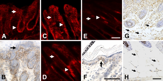
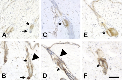
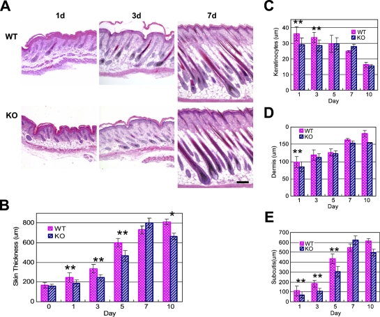
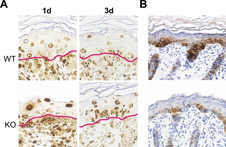
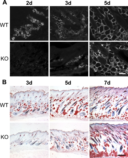
In early postnatal mouse skin, the NG2 proteoglycan is expressed in the subcutis, the dermis, the outer root sheath of hair follicles, and the basal keratinocyte layer of the epidermis. With further development, NG2 is most prominently expressed by stem cells in the hair follicle bulge region, as also observed in adult human skin. During telogen and anagen phases of the adult hair cycle, NG2 is also found in stem cell populations that reside in dermal papillae and the outer root sheaths of hair follicles. Ablation of NG2 produces alterations in both the epidermis and subcutis layers of neonatal skin. Compared with wild type, the NG2 null epidermis does not achieve its full thickness due to reduced proliferation of basal keratinocytes that serve as the stem cell population in this layer. Thickening of the subcutis is also delayed in NG2 null skin due to deficiencies in the adipocyte population.


Figures

Figure 1
Figure 1
NG2 expression during early postnatal skin development. On postnatal day 1 (A), NG2 expression is strong in both the dermis (arrow) and outer root sheath of the hair follicle (arrowhead). Superficial to the dermis, immunoperoxidase staining reveals weaker expression of NG2 in the basal layer of keratinocytes (arrow in B). At day 3 (C), NG2 immunoreactivity remains strong in the hair follicle outer root sheath (arrowhead) but begins to diminish in the dermis (arrow). By days 7 (D) and 10 (E), NG2 has virtually disappeared from the dermis (arrow) and is mainly seen in the hair follicle bulge region (arrowheads). Immunoperoxidase labeling (F) reveals NG2 expression by basal keratinocytes at day 10 (arrow). In situ hybridization was used at postnatal days 1 (G) and 7 (H) to detect NG2 transcripts in adipocytes located in the subcutis (arrows); i.e., beneath the dermis and hair follicles. Bar = 50 μm.
Figure 2
Figure 2
NG2 expression by stem cells as a function of the hair cycle. Cytokeratin (CK)-15 immunoreactivity (A,C,E) was used to mark stem cells in the bulge region during telogen (A,B), anagen (C,D), and catagen (E,F) phases of the hair cycle. NG2 expression (B,D,F) is always seen in the bulge region (asterisks), but during telogen and anagen it is also seen in dermal papillae (arrows in telogen A,B) and the outer root sheath of hair follicles (arrowheads). Bar = 100 μm.
Figure 3
Figure 3
Skin development in the NG2 null mouse. Images of hematoxylin- and eosin-stained skin sections (transverse) were prepared from wild-type (WT) and NG2 null mice during the first 10 days postnatally (A) and used to measure the overall skin thickness (B) and the thickness of the epidermis (C), dermis (D), and subcutis (E). At least six mice were used at each time point to establish the average values shown in the panels. Results were analyzed using the two-tailed Student's t-test. Statistically significant differences between WT and NG2 null samples are designated as follows: **p<0.01; *p<0.05. Bar = 200 μm.
Figure 4
Figure 4
Decreased keratinocyte production in the NG2 null mouse. (A) Following BrdU administration at 17 days of gestation, BrdU-labeled cells in the skin were visualized at days 1 and 3 by staining with anti-BrdU antibody. Red lines in each panel mark the dermis/epidermis boundary. (B) Immunoperoxidase labeling for CK-5 illustrates the deficiency in basal keratinocytes in NG2 null skin.
Figure 5
Figure 5
Altered adipocyte development in the NG2 null mouse. (A) Sections of WT and NG2 null skin at days 2, 3, and 5 were immunostained with antibody against aP2/FABP-4. Expression of aP2/FABP-4 is delayed in NG2 null adipocytes. (B) Staining with Oil Red O was used to examine lipid deposition in WT and NG2 null skin at days 3, 5, and 7. Delay in lipid deposition mirrors the delay in FABP-4 expression shown in A. Bars: A = 100 μm; B = 200 μm.

Similar articles

Cited by

References

    1. Beer H, Bittner M, Niklaus G, Munding C, Max N, Goppelt A, Werner S (2005) The fibroblast growth factor binding protein is a novel interaction partner of FGF-7, FGF-10, and FGF-22 and regulates FGF activity: implications for epithelial repair. Oncogene 24:5269–5277 - PubMed
    1. Braun S, Krampert M, Bodó E, Kümin A, Born-Berclaz, Paus R, Werner S (2006) Keratinocyte growth factor protects epidermis and hair follicles from cell death induced by UV radiation, chemotherapeutic or toxic agents. J Cell Sci 119:4841–4849 - PubMed
    1. Burg M, Nishiyama A, Stallcup WB (1997) A central segment of the NG2 proteoglycan is critical for the ability of glioma cells to bind and migrate toward type VI collagen. Exp Cell Res 235:254–264 - PubMed
    1. Cheng B, Liu H, Fu X, Sun T, Sheng Z (2007) Recombinant human platelet-derived growth factor enhanced dermal wound healing by a pathway involving ERK and c-fos in diabetic rats. J Dermatol Sci 45:193–201. Published online January 31, 2007 (DOI: 10.1016/J.Jdermsci.2006.11.014) - PubMed
    1. Commo S, Gaillard O, Bernard B (2000) The human hair follicle contains two distinct K19 positive compartments in the outer root sheath: a unifying hypothesis for stem cell reservoir. Differentiation 66:157–164 - PubMed

Publication types

MeSH terms