

Tactile-associated fMRI recruitment of the cervical cord in healthy subjects
- PMID: 18041739
- PMCID: PMC6870728
- DOI: 10.1002/hbm.20499
Tactile-associated fMRI recruitment of the cervical cord in healthy subjects
Abstract
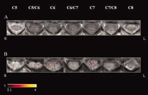
Using spinal cord functional magnetic resonance imaging (fMRI), 12 right-handed healthy subjects were scanned during a tactile stimulation of the palm of the right hand. The task-related mean signal change was computed for all activated voxels within the cervical cord, and separately, in the four cord quadrants (right and left anterior, right and left posterior) from C5 to C8. The frequency of fMRI activity at each cord level was obtained by assigning a score of 25% at each active quadrant and by averaging the percentage of active quadrants at each level of all subjects. The difference in the occurrence of fMRI activity (a) in right versus left, and anterior versus posterior cord, and (b) among the different cord levels, was evaluated using a random effect logistic regression model, with the frequency of fMRI activity as the dependent variable and the subject as the grouping factor. The task-related mean signal change of all activated voxels of the cord was 3.2% (SD = 0.8%). During the tactile stimulation, subjects showed a higher occurrence of fMRI cord activity in the right than in the left cervical cord (odds ratio = 2.25, 95% confidence interval = 1.31-3.87, P = 0.003). A significant heterogeneity in frequency of fMRI activity between cord levels was also observed (P < 0.001), with the highest frequencies of fMRI activity detected at C6 and C7. Spinal cord fMRI enables to obtain reliable physiological information on the activity of human spinal circuits associated to tactile stimulation. This holds significant promise for a better planning and conduct of studies of people with diseased spinal cords.
(c) 2007 Wiley-Liss, Inc.
Figures
References
-
- Brodal A ( 1981): Neurological Anatomy in Relation to Clinical Medicine. New York: Oxford University Press.
-
- Brown AG ( 1981): Organization of the Spinal Cord. New York: Springer Verlag.
-
- Constable RT,Gore JC ( 1992): The loss of small objects in variable TE imaging: Implications for FSE, RARE and EPI. Magn Reson Med 28: 9–24. - PubMed
-
- Friston KJ,Jezzard P,Turner R ( 1994): Analysis of functional MRI time series. Hum Brain Mapp 1: 153–171.
-
- Fujita H,Meyer E,Reutens DC,Kuwabara H,Evans AC,Gjedde A ( 1997): Cerebral [15O] water clearance in humans determined by positron emission tomography. II. Vascular responses to vibrotactile stimulation. J Cereb Blood Flow Metab 17: 73–79. - PubMed
Publication types
MeSH terms
LinkOut - more resources
Full Text Sources
Miscellaneous
