

Elevated activity and microglial expression of myeloperoxidase in demyelinated cerebral cortex in multiple sclerosis
- PMID: 18042261
- PMCID: PMC8095620
- DOI: 10.1111/j.1750-3639.2007.00110.x
Elevated activity and microglial expression of myeloperoxidase in demyelinated cerebral cortex in multiple sclerosis
Abstract
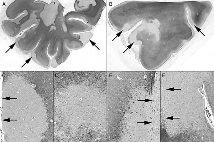
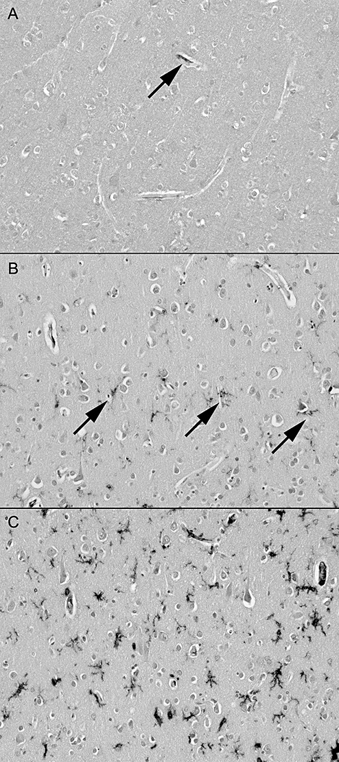
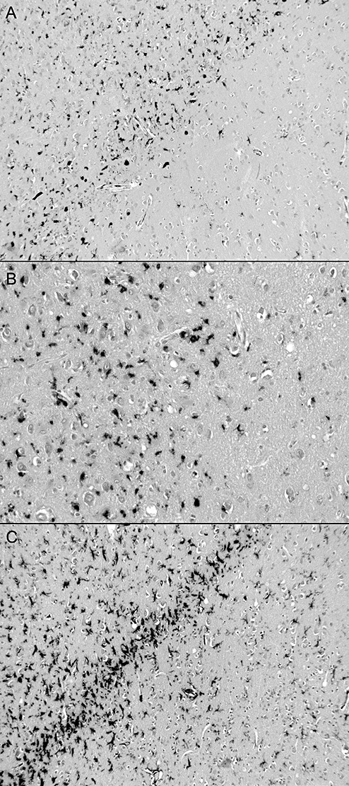
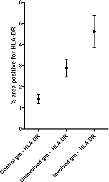
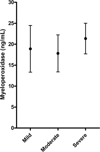
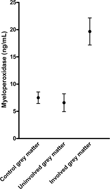
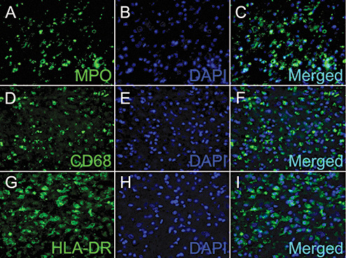
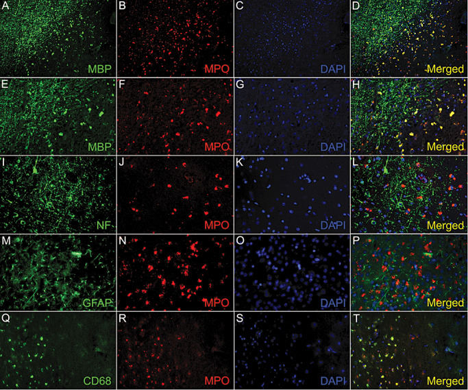
Recent studies have revealed extensive cortical demyelination in patients with progressive multiple sclerosis (MS). Demyelination in gray matter lesions is associated with activation of microglia. Macrophages and microglia are known to express myeloperoxidase (MPO) and generate reactive oxygen species during myelin phagocytosis in the white matter. In the present study we examined the extent of microglial activation in the cerebral cortex and the relationship of microglial activation and MPO activity to cortical demyelination. Twenty-one cases of neuropathologically confirmed multiple sclerosis, with 34 cortical lesions, were used to assess microglial activation. HLA-DR immunolabeling of activated microglia was significantly higher in demyelinated MS cortex than control cortex and, within the MS cohort, was significantly greater within cortical lesions than in matched non-demyelinated areas of cortex. In homogenates of MS cortex, cortical demyelination was associated with significantly elevated MPO activity. Immunohistochemistry revealed MPO in CD68-positive microglia within cortical plaques, particularly toward the edge of the plaques, but not in microglia in adjacent non-demyelinated cortex. Cortical demyelination in MS is associated with increased activity of MPO, which is expressed by a CD68-positive subset of activated microglia, suggesting that microglial production of reactive oxygen species is likely to be involved in cortical demyelination.
Figures
Similar articles
-
Elevated myeloperoxidase activity in white matter in multiple sclerosis.Neurosci Lett. 2008 Oct 24;444(2):195-8. doi: 10.1016/j.neulet.2008.08.035. Epub 2008 Aug 15. Neurosci Lett. 2008. PMID: 18723077
-
Meningeal inflammation and cortical demyelination in acute multiple sclerosis.Ann Neurol. 2018 Dec;84(6):829-842. doi: 10.1002/ana.25365. Epub 2018 Nov 30. Ann Neurol. 2018. PMID: 30362156
-
Cortical neuronal densities and cerebral white matter demyelination in multiple sclerosis: a retrospective study.Lancet Neurol. 2018 Oct;17(10):870-884. doi: 10.1016/S1474-4422(18)30245-X. Epub 2018 Aug 22. Lancet Neurol. 2018. PMID: 30143361 Free PMC article.
-
Grey matter pathology in multiple sclerosis.Acta Neurol Scand Suppl. 2006;183:48-50. doi: 10.1111/j.1600-0404.2006.00615.x. Acta Neurol Scand Suppl. 2006. PMID: 16637929 Review.
-
Gray matter pathology and multiple sclerosis.Curr Neurol Neurosci Rep. 2009 Sep;9(5):399-404. doi: 10.1007/s11910-009-0058-x. Curr Neurol Neurosci Rep. 2009. PMID: 19664370 Review.
Cited by
-
Myeloperoxidase Molecular MRI Reveals Synergistic Combination Therapy in Murine Experimental Autoimmune Neuroinflammation.Radiology. 2019 Oct;293(1):158-165. doi: 10.1148/radiol.2019182492. Epub 2019 Sep 3. Radiology. 2019. PMID: 31478802 Free PMC article.
-
Effects and mechanism of myeloperoxidase on microglia in the early stage of intracerebral hemorrhage.Front Neurosci. 2022 Dec 9;16:1046244. doi: 10.3389/fnins.2022.1046244. eCollection 2022. Front Neurosci. 2022. PMID: 36570834 Free PMC article.
-
Myeloperoxidase exerts anti-tumor activity in glioma after radiotherapy.Neoplasia. 2022 Apr;26:100779. doi: 10.1016/j.neo.2022.100779. Epub 2022 Mar 2. Neoplasia. 2022. PMID: 35247801 Free PMC article.
-
TRPA1 Role in Inflammatory Disorders: What Is Known So Far?Int J Mol Sci. 2022 Apr 20;23(9):4529. doi: 10.3390/ijms23094529. Int J Mol Sci. 2022. PMID: 35562920 Free PMC article. Review.
-
Manual Segmentation of MS Cortical Lesions Using MRI: A Comparison of 3 MRI Reading Protocols.AJNR Am J Neuroradiol. 2016 Sep;37(9):1623-8. doi: 10.3174/ajnr.A4799. Epub 2016 May 19. AJNR Am J Neuroradiol. 2016. PMID: 27197988 Free PMC article.
References
-
- Bo L, Vedeler CA, Nyland H, Trapp BD, Mork SJ (2003) Intracortical multiple sclerosis lesions are not associated with increased lymphocyte infiltration. Mult Scler 9:323–331. - PubMed
-
- Bo L, Vedeler CA, Nyland HI, Trapp BD, Mork SJ (2003) Subpial demyelination in the cerebral cortex of multiple sclerosis patients. J Neuropathol Exp Neurol 62:723–732. - PubMed
Publication types
MeSH terms
Substances
Grants and funding
LinkOut - more resources
Full Text Sources
Other Literature Sources
Medical
Research Materials
Miscellaneous
