. 2007 Nov 28:8:436.
doi: 10.1186/1471-2164-8-436.

Characterization of Smoc-1 uncovers two transcript variants showing differential tissue and age specific expression in Bubalus bubalis

Affiliations

Characterization of Smoc-1 uncovers two transcript variants showing differential tissue and age specific expression in Bubalus bubalis

Jyoti Srivastava et al. BMC Genomics. .

Abstract

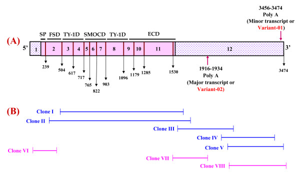
Background: Secreted modular calcium binding protein-1 (Smoc-1) belongs to the BM-40 family which has been implicated with tissue remodeling, angiogenesis and bone mineralization. Besides its anticipated role in embryogenesis, Smoc-1 has been characterized only in a few mammalian species. We made use of the consensus sequence (5' CACCTCTCCACCTGCC 3') of 33.15 repeat loci to explore the buffalo transcriptome and uncovered the Smoc-1 transcript tagged with this repeat. The main objective of this study was to gain an insight into its structural and functional organization, and expressional status of Smoc-1 in water buffalo, Bubalus bubalis.

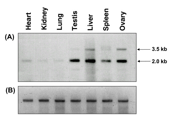
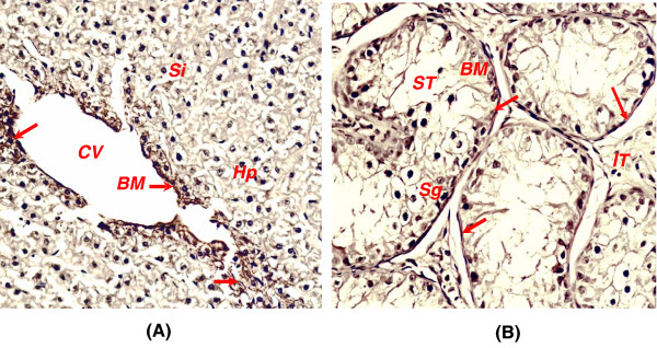
Results: We cloned and characterized the buffalo Smoc-1, including its copy number status, in-vitro protein expression, tissue & age specific transcription/translation, chromosomal mapping and localization to the basement membrane zone. Buffalo Smoc-1 was found to encode a secreted matricellular glycoprotein containing two EF-hand calcium binding motifs homologous to that of BM-40/SPARC family. In buffalo, this single copy gene consisted of 12 exons and was mapped onto the acrocentric chromosome 11. Though this gene was found to be evolutionarily conserved, the buffalo Smoc-1 showed conspicuous nucleotide/amino acid changes altering its secondary structure compared to that in other mammals. In silico analysis of the Smoc-1 proposed its glycoprotein nature with a calcium dependent conformation. Further, we unveiled two transcript variants of this gene, varying in their 3'UTR lengths but both coding for identical protein(s). Smoc-1 evinced highest expression of both the variants in liver and modest to negligible in other tissues. The relative expression of variant-02 was markedly higher compared to that of variant-01 in all the tissues examined. Moreover, expression of Smoc-1, though modest during the early ages, was conspicuously enhanced after 1 year and remained consistently higher during the entire life span of buffalo with gradual increment in expression of variant-02. Immunohistochemically, Smoc-1 was localized in the basement membrane zones and extracellular matrices of various tissues.

Conclusion: These data added to our understandings about the tissue, age and species specific functions of the Smoc-1. It also enabled us to demonstrate varying expression of the two transcript variants of Smoc-1 amongst different somatic tissues/gonads and ages, in spite of their identical coding frames. Pursuance of these variants for their roles in various disease phenotypes such as hepatocellular carcinoma and angiogenesis is envisaged to establish broader biological significance of this gene.


Figures

Figure 1
Figure 1
Diagrammatic illustration showing cloning strategy of buffalo Smoc-1. Smoc-1 structure representing 5'/3'UTRs, domain organization and nucleotide boundary of each exon is shown in (A). The strategy for isolation of the Smoc-1 is given in (B). Different fragments generated by end point PCR (blue) and RACE (pink) used to deduce the full length CDS are shown along with their nucleotide boundaries. Clone I covered nucleotides 318–1580; clone II, 119–1603; clone III, 1461–2473; clone IV, 2307–3328; and clone V, 2435–3428; clone VI, 1–649; clone VII, 1407–1915 and clone VIII, 2435–3474. Two transcript variants of Smoc-1 with their 3'UTR length variation are shown. Poly(A) tails for both the variants, -01 (3474 bp) and -02 (1934 bp) are marked by arrows in 'A'.
Figure 2
Figure 2
Two transcript variants of Smoc-1. Northern blot showing two transcript variants of Smoc-1 in different somatic and gonadal tissues of water buffalo. Note two distinct bands with varying intensity in each tissue along with highest expression in liver, and lowest in lung, kidney, and heart.
Figure 3
Figure 3
Structure-based alignment of the Smoc-1 protein from different mammalian species. The sequences were aligned across the species for FS (A), TY1-1 (B), Smoc-1 (C), TY1-2 (D) and EC (E) domains. Mutational hotspots in buffalo Smoc-1 are shown red boldface. Most of the observed changes in buffalo Smoc-1 were shared either by cattle or human. The "→" indicates the potential N-glycosylation site. Pairs of numbers above the sequence correspond to cysteines indicating the predicted disulphide bonds based on the disulphide linkage of BM-40 and thyroglobulin. Both EF hand motifs are underlined and calcium coordinating residues are overshadowed grey. Conserved amino acids are indicated by the stars below and cysteines with a black background.
Figure 4
Figure 4
Chromosomal mapping of Smoc-1 gene at chromosome 11 in buffalo. Fluorescence in situ hybridization demonstrating the presence of Smoc-1 gene on the distal arm of the acrocentric chromosome 11 (A) and detailed mapping of this gene with respect to its position on the G-banded ideogram following the chromosome nomenclature standardized by ISCNDB, 2000 (B).
Figure 5
Figure 5
Highest expression of Smoc-1 in liver. RT-PCR showed expression in liver only but Southern hybridization detected reduced signals in testis, ovary and spleen (A) whereas control RT-PCR with β-actin showed almost equal intensity signal in each tissue (B). Quantitative expression of Smoc-1 based on Real Time PCR confirmed maximum expression (163–364 folds) in liver in five different animals compared to that in lung used as the calibrator (C). Note the buffalo spermatozoa from four different animals (BS1-4) also showed 4–7 folds transcripts in four different animals, similar to that in testis. Relative quantitation also demonstrated higher expression (1.2–3.5 times) of variant-02 compared to that of -01 in each of the tissues examined (D). Western blotting with anti-SySmoc1-pAb (E) and anti-β-actin-mAb as positive control (F) substantiated the highest expression of Smoc-1 protein in liver. The quantitative expression carried out using cDNA isolated from blood lymphocytes of different age group of animals is shown in (G). Note the markedly increased expression of Smoc-1 in animals 10 months and beyond. Relative quantitation showing higher expression of variant-01 in the animal up to ~6 months of age and that of variant-02 in the animals of age 6 months and beyond (H).
Figure 6
Figure 6
Indirect Immunohistochemistry of buffalo tissue sections using Anti-SySmoc-1-pAb. Distribution of Smoc-1 in basement membrane zone of endothelial cell layer and extracellular matrices of liver (A) and specific expression around basement membrane zone of the tubuli seminiferi in the testis (B). Note the localization of Smoc-1 protein in the basement membrane zones indicated by red arrows. The bars represent 5 μm in panels A-B. Hp denotes hepatocytes; Si, sinusoids; CV, central vein and Em, discontinuous endothelial cell of central vein in 'A', and ST, seminiferous tubules; BM, basement membrane zone; Sg, Spermatogonia and IT, interstitial tissues in 'B'.

Similar articles

Cited by

References

    1. Bornstein P, Sage EH. Matricellular proteins, extracellular modulators of cell function. Curr Opin Cell Biol. 2002;14:608–616. doi: 10.1016/S0955-0674(02)00361-7. - DOI - PubMed
    1. Termine JD, Kleinman HK, Whitson SW, Conn KM, McGarvey ML, Martin GR. Osteonectin, a bone-specific protein linking mineral to collagen. Cell. 1981;26:99–105. doi: 10.1016/0092-8674(81)90037-4. - DOI - PubMed
    1. Lane TF, Sage EH. The biology of SPARC, a protein that modulates cell-matrix interactions. FASEB J. 1994;8:163–173. - PubMed
    1. Brekken RA, Sage EH. SPARC, a matricellular protein, at the crossroads of cell-matrix communication. Matrix Biol. 2001;19:816–827. doi: 10.1016/S0945-053X(00)00133-5. - DOI - PubMed
    1. Ledda MF, Adris S, Bravo AI, Kairiyama C, Bover L, Chernajovsky Y, Mordoh J, Podhajcer OL. Supression of SPARC expression by the antisensce RNA abrogates the tumorogenicity of human melanoma cells. Nat Med. 1997;3:171–176. doi: 10.1038/nm0297-171. - DOI - PubMed

Publication types

MeSH terms

Associated data