. 2007 Nov 30;28(4):614-23.
doi: 10.1016/j.molcel.2007.09.013.

Structure of the Pho85-Pho80 CDK-cyclin complex of the phosphate-responsive signal transduction pathway

Affiliations

Structure of the Pho85-Pho80 CDK-cyclin complex of the phosphate-responsive signal transduction pathway

Kexin Huang et al. Mol Cell. .

Abstract

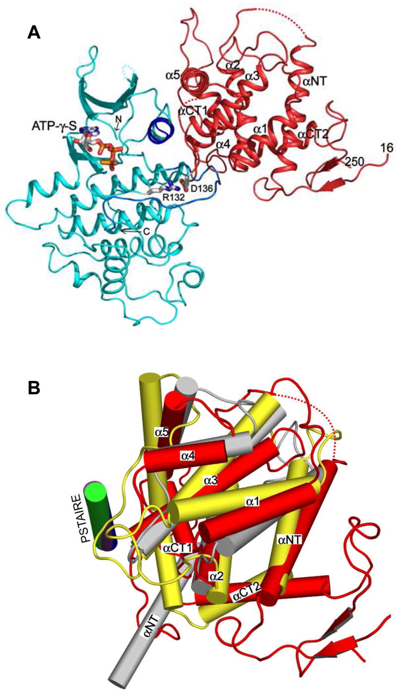
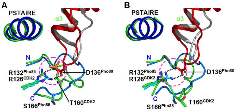
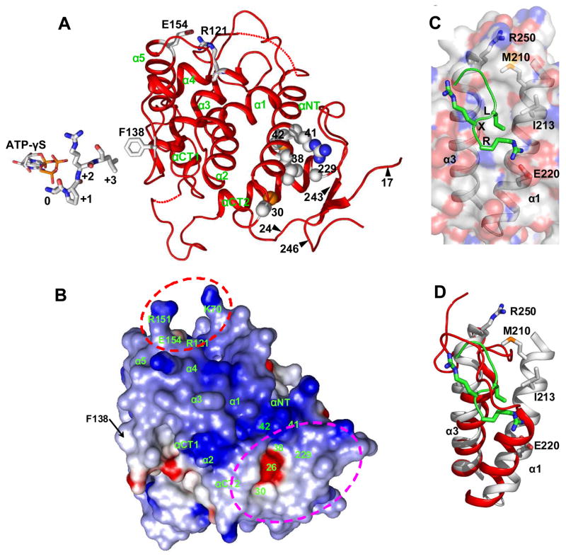
The ability to sense and respond appropriately to environmental changes is a primary requirement of all living organisms. In response to phosphate limitation, Saccharomyces cerevisiae induces transcription of a set of genes involved in the regulation of phosphate acquisition from the ambient environment. A signal transduction pathway (the PHO pathway) mediates this response, with Pho85-Pho80 playing a vital role. Here we report the X-ray structure of Pho85-Pho80, a prototypic structure of a CDK-cyclin complex functioning in transcriptional regulation in response to environmental changes. The structure revealed a specific salt link between a Pho85 arginine and a Pho80 aspartate that makes phosphorylation of the Pho85 activation loop dispensable and that maintains a Pho80 loop conformation for possible substrate recognition. It further showed two sites on the Pho80 cyclin for high-affinity binding of the transcription factor substrate (Pho4) and the CDK inhibitor (Pho81) that are markedly distant to each other and the active site.


Figures

Figure 1
Figure 1. Structure of Pho85-Pho80
(A) Ribbon backbone trace of the crystal structure of the Pho85-Pho80 complex with the bound ATP analog, ATP-γ-S. Pho85 is shown in cyan, and its PSTAIRE helix and activation loop in blue and marine, respectively. Pho80 is depicted in red. The ATP-γ-S is represented in ball-and-stick model. The amino- and carboxy-terminal ends of Pho85 are marked by N and C, respectively, and those of Pho80 are denoted by residue numbers 16 and 250, respectively. The first 15 and last 43 residues in Pho80 are not visible (or disordered) in the electron density map, indicating their flexibilities. The long loops in Pho80 include the visible N-terminal 17- and C-terminal 22-residue loops, the 25-residue loop between αNT and α1, and the 33-residue loop between αCT1 and αCT2 helices (see Supplementary Figure S1). The two dotted lines in the Pho80 trace represent disordered segments (see Supplementary Figure S1). R132 on Pho85 and D136 on Pho80 form a salt link (details shown in Figure 2). (B) Relative positions of Pho80 (red), cyclin A (grey) and p25 (yellow) following superposition of their cognate kinase subunits as they occur in the structures of the Pho85-Pho80-ATP-γ-S complex, the phosphorylated CDK2-cyclin A complex (Brown et al., 1999) and CDK5-p25 (Tarricone et al., 2001). The orientation of Pho80 is identical to that shown in panel A. The depiction of the α-helices by cylinders emphasizes the differences between the relative orientations of the cyclins. p25 lacks the equivalent of the 2-turn α4 helices in Pho80 and cyclin A, and in its place is a turn which includes the P247 residue. The PSTAIRE (Pho85 and CDK2) or PSAALRE (p25) helices are shown in blue (Pho85), green (CDK2) and purple (CDK5). Although the PSTAIRE/PSSALRE helices are completely superimposable, the contacts they make with their cognate cyclins vary (e.g., see Figures S2 and S3). In the truncated cyclin A structure, only the first helix (αNT) and the first cyclin box are shown. The succeeding two helices (α1′ and α2′) in cyclin A (not shown) constitute the first two helices of the second cyclin box and adopt positions different from those of αCT1 and αCT2, respectively, in Pho80 or p25. The αNT helix of cyclin A makes significant interaction with the C-terminal lobe of CDK2 (Russo et al., 1996b). In contrast, the αNT helices of Pho80 and p25, which approximately adopt similar positions, do not participate in kinase binding as they lie on the opposite side of the cyclin structures to the kinase-cyclin interface or the location of αNT in cyclin A.
Figure 2
Figure 2. A Salt Link Between Pho85 R132 and Pho80 D136 Which Makes T-loop Phosphorylation Dispensable
(A) Relative positions of the loop between α3 and α4 in Pho80 (red) and cyclin A (grey) and the PSTAIRE helices and segments of the activation loops in Pho85 (residues 155 – 168) (marine) and phosphorylated (T160) CDK2 (residues 149 – 163) (green). The overlay is based on the superposition of the structures of the kinase subunits as they occur in the structures of the Pho85-Pho80-ATP-γ-S complex, and the phosphorylated CDK2-cyclin A complex (Brown et al., 1999). The α3-α4 loop in Pho80 deploys D136, hence the “D-loop” name. The activation loop T160 (with the phosphoryl adduct) of CDK2 corresponds to S166 in Pho85. Pho85 R132 and CDK2 R126 are superimposable. CDK5 R125 adopts almost identical position as those of Pho85 R132 and CDK2 R126 (data not shown). Pho80 D136 has no counterpart in cyclin A and p25. A more global view of the D-loop and the salt link is shown in Figure 1A. (B) Similar superposition as shown in panel A, except that the CDK2 structure is the unphosphorylated form (Jeffrey et al., 1995). The position of R126 of CDK2 is essentially unchanged from that in the phosphorylated CDK2 (panel A). Note the significant change in the conformation of the segment of the T-loop of CDK2 following phosphorylation (compare with that in panel A).
Figure 3
Figure 3. Three Functional Sites of the Pho80 Cyclin Subunit
(A) Ribbon trace of Pho80. The regions of the three functional sites are identified as follows (see also text): The bound ATP-γ-S (stick representation) marking the location of the kinase active site region; the cluster of residues C30, L38, R41, M42 and G229 and segments of residues 17 to 24 and 243 to 246 forming a large cavity for possible tight interaction with the site on Pho4 distal to the phosphorylatable sites; and the salt link between R121 and E154 in a region for docking the inhibitor, Pho81. The peptide near the ATP-γ-S corresponds to a model of the the SPRL consensus sequence of the five Pho4 phosphorylations sites (O’Neill et al., 1996). Placement of the peptide was derived by superimposing the atomic coordinates of only the Pho85 and CDK2 subunits in the Pho85-Pho80-ATP-γ-S structure and the structure of the phosphorylated CDK2-cyclin A with a bound peptide containing the SPRK consensus sequence and AMPPNP-Mg2+ (Brown et al., 1999). Changing the lysine at the +3 position to leucine to conform to the Pho4 consensus sequence, SPXI/L, indicates that the leucine sidechain would be within van der Waals distance to the F138 located on the α3-α4 (or D-) loop in Pho80. (B) Electrostatic surface potential surface of Pho80 contoured at −10kT (red) and +10kT (blue) calculated with GRASP (Nicholls et al., 1991). Pho80 is in identical orientation as in panel A. The region involved in binding of the CKI Pho81 is enclosed by red dashed lines. The region involved in docking of the site on Pho4 distal to the phosphorylation sites is enclosed in magenta dashed lines. (C) Hydrophobic patch on cyclin A for docking the RXL motif of substrates or CDK inhibitors. The bound ligand, with backbone trace in green, shows only the RRL sequence (the equivalent of the RXL motif) of the substrate recruitment peptide RRLFGE of p107 (Brown et al., 1999). The residues M210, I213 and E220 on α1 and R250 on α3 define the specificity of the patch in a group of cell cycle cyclins for the RXL motif (Endicott et al., 1999). (D) Identical to panel C, but with the surface of cyclin A removed for clarity, and the α1 and α3 helices and their preceding loops of Pho80 superimposed. The first two turns of α1 of cyclin A, which provides M210 and I213, are missing in the corresponding α1 of Pho80.

References

    1. Adams PD, Sellers WR, Sharma SK, Wu AD, Nalin CM, Kaelin WG., Jr Identification of a cyclin-cdk2 recognition motif present in substrates and p21-like cyclin-dependent kinase inhibitors. Mol Cell Biol. 1996;16:6623–6633. - PMC - PubMed
    1. Brown NR, Noble ME, Endicott JA, Johnson LN. The structural basis for specificity of substrate and recruitment peptides for cyclin-dependent kinases. Nature Cell Biol. 1999;1:438–443. - PubMed
    1. Byrne M, Miller N, Springer M, O’Shea EK. A distal, high-affinity binding site on the cyclin-CDK substrate Pho4 is important for its phosphorylation and regulation. J Mol Biol. 2004;335:57–70. - PubMed
    1. Carroll AS, O’Shea EK. Pho85 and signaling environmental conditions. Trends Biochem Sci. 2002;27:87–93. - PubMed
    1. Endicott JA, Noble ME, Tucker JA. Cyclin-dependent kinases: inhibition and substrate recognition. Curr Opin Struct Biol. 1999;9:738–744. - PubMed

Publication types

MeSH terms