. 2007 Nov 30;28(4):638-51.
doi: 10.1016/j.molcel.2007.11.001.

Sae2 is an endonuclease that processes hairpin DNA cooperatively with the Mre11/Rad50/Xrs2 complex

Affiliations

Sae2 is an endonuclease that processes hairpin DNA cooperatively with the Mre11/Rad50/Xrs2 complex

Bettina M Lengsfeld et al. Mol Cell. .

Abstract

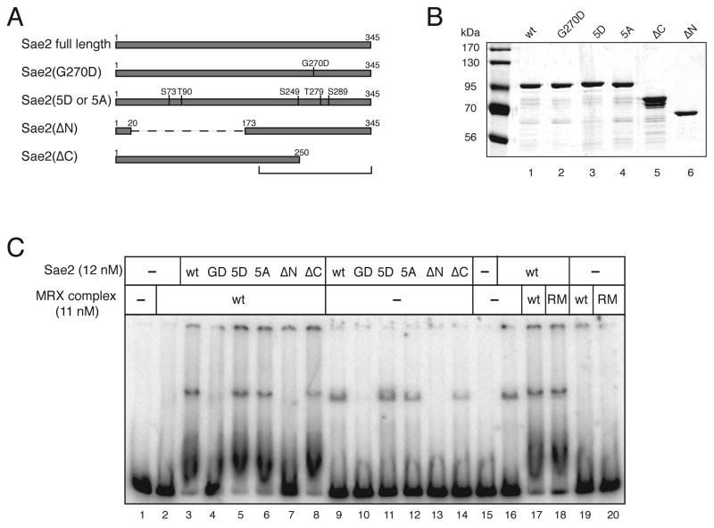
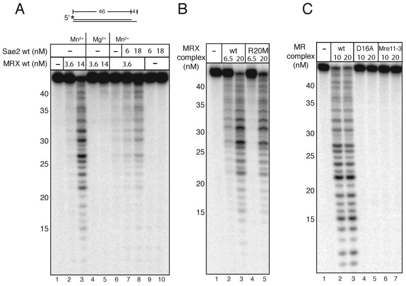
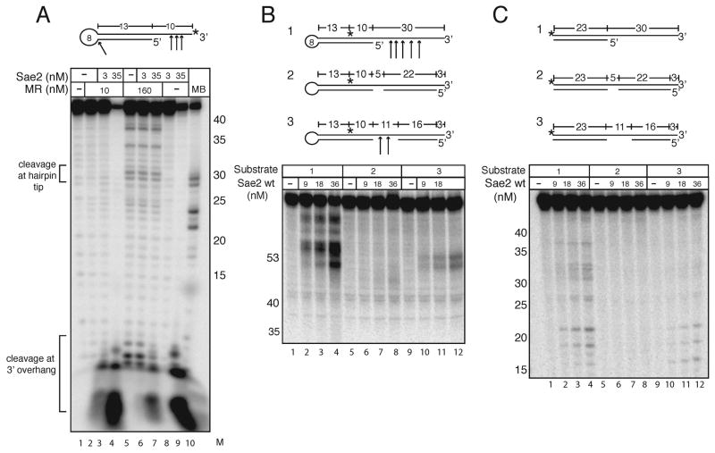
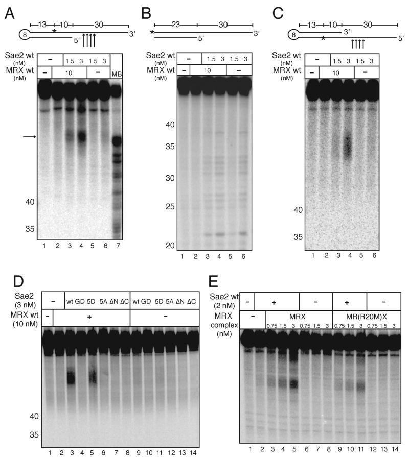
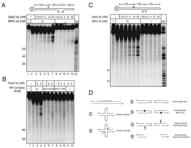
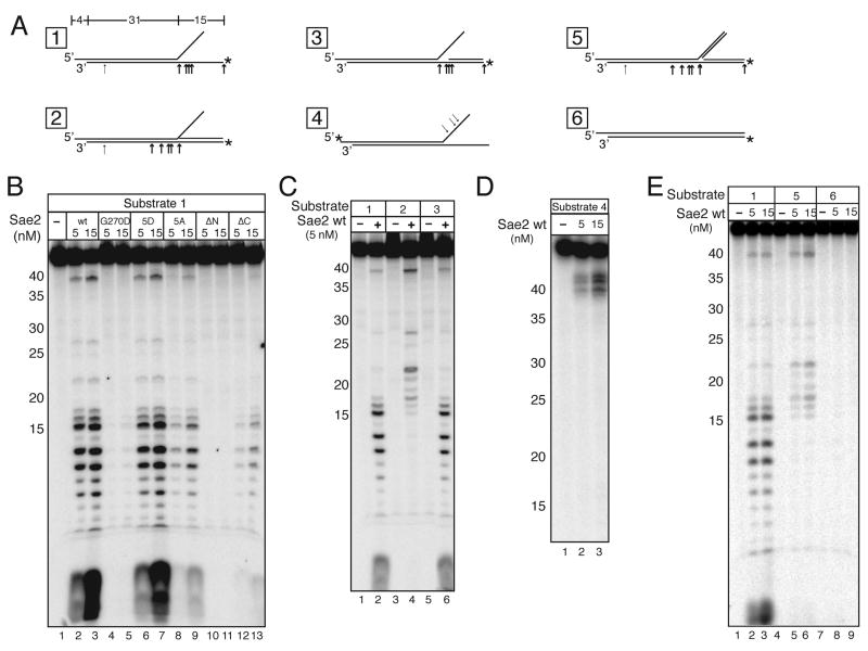
Mre11/Rad50 complexes in all organisms function in the repair of DNA double-strand breaks. In budding yeast, genetic evidence suggests that the Sae2 protein is essential for the processing of hairpin DNA intermediates and meiotic double-strand breaks by Mre11/Rad50 complexes, but the biochemical basis of this functional relationship is not known. Here we demonstrate that recombinant Sae2 binds DNA and exhibits endonuclease activity on single-stranded DNA independently of Mre11/Rad50 complexes, but hairpin DNA structures are cleaved cooperatively in the presence of Mre11/Rad50 or Mre11/Rad50/Xrs2. Hairpin structures are not processed at the tip by Sae2 but rather at single-stranded DNA regions adjacent to the hairpin. Truncation and missense mutants of Sae2 inactivate this endonuclease activity in vitro and fail to complement Deltasae2 strains in vivo for meiosis and recombination involving hairpin intermediates, suggesting that the catalytic activities of Sae2 are important for its biological functions.


Figures

Figure 1
Figure 1
Expression and DNA binding activity of recombinant Sae2. (A) Schematic representation of full-length wild-type Sae2, and mutants G270D, 5D (residues indicated changed to aspartate), 5A (residues indicated changed to alanine), ΔN (Δ a.a. 21 to 173) and ΔC (Δ a.a. 251 to 345). (B) SDS-PAGE of purified Sae2 wild-type and mutant proteins, approximately 100 ng total protein each. (C) Wild-type (wt) and mutant Sae2 proteins were incubated with a 249 bp double-stranded DNA substrate and analyzed in a 8% native polyacrylamide gel in the presence of wild-type (wt) or R20M (RM) MRX complex. Protein-DNA complex 1 and complex 2 are indicated (described in text).
Figure 2
Figure 2
Nuclease activities of MR and MRX complexes. (A) MRX was incubated with a 5′ [32P]-labeled 46 bp DNA substrate containing a 4 nt recessed 3′ end in 1 mM MnCl2 or 5 mM MgCl2 with wild-type Sae2 protein as indicated. (B) The Rad50S MR(R20M)X complex was assayed in comparison to the wild-type complex as in (A) in 1 mM MnCl2. (C) The nuclease-deficient complexes M(D16A)R and M(Mre11-3)R were assayed in comparison to the wild-type MR complex as in (A) in 1 mM MnCl2.
Figure 3
Figure 3
Sae2 cleaves hairpin DNA. (A) Wild-type MR and Sae2 were incubated with a 3′ [32P]-labeled hairpin DNA substrate with 1 mM MnCl2 and 0.5 mM ATP as indicated and separated in a denaturing polyacrylamide gel. Substrate was also incubated with Mung Bean nuclease as a control to show the location of hairpin cut at the tip (MB). Numbers in the “M” lane indicate the positions of DNA standards run in the same gel. (B) Reactions were performed as in (A) with Sae2 only, 5 mM MgCl2, and internally-labeled hairpin substrates as shown. (C) Reactions were performed with substrates identical to those in (B) except lacking the hairpin loops.
Figure 4
Figure 4
Sae2 and MRX cooperatively cleave hairpin structures. (A) Wild-type MRX and Sae2 were incubated in 5 mM MgCl2 with an internally [32P]-labeled hairpin DNA substrate as in Fig. 3B. The primary cleavage products (arrow) are not cut at the tip of the hairpin but in the single-stranded overhang as shown in the diagram. MB control as in Fig. 3. (B) Reactions were performed as in (A) but with substrates lacking the hairpin loop. (C) Wild-type MRX and Sae2 were incubated in 5 mM MgCl2 with an internally [32P]-labeled hairpin DNA substrate with the opposite polarity compared to the substrate shown in (A). (D) Wild-type MRX and wild-type and mutant Sae2 proteins were incubated in 5 mM MgCl2 with an internally [32P]-labeled hairpin DNA substrate as in (B). (E) Wild-type Sae2 and wild-type MRX and Rad50S MR(R20M)X complexes were assayed as in (A).
Figure 5
Figure 5
Mre11 exonuclease activity facilitates Sae2 hairpin removal. (A) An internally [32P]-labeled double hairpin DNA substrate was incubated first with wild-type MRX in 1 mM MnCl2 for 20 minutes; Sae2 was then added with 5 mM MgCl2 and the reactions were continued for an additional 30 minutes before separation on a denaturing polyacrylamide gel. MB control as in Fig. 3. (B) Reactions were performed as in (A) but with wild-type MR and Mre11 nuclease-deficient complexes M(D16A)R and M(Mre11-3)R as indicated. (C) Hairpin cleavage assays as in (A) except that the substrate has a nick instead of a gap. (D) Model of MRX/Sae2 hairpin processing. Dotted arrow represents predicted MRX/Sae2-independent cleavage of the cruciform; Pacman represents MRX 3′ to 5′ exonuclease activity; bold arrow represents Sae2 cleavage. The location of the cruciform cleavage site is assumed to be at the base of the cruciform as shown but this has not been demonstrated in vivo.
Figure 6
Figure 6
Sae2 cleaves single-stranded DNA in branched DNA structures. (A) Diagram of DNA substrates 1 to 4 with locations of [32P] labels (asterisks) and summary of predominant cleavage sites (arrows) as shown. (B) Wild-type and mutant Sae2 proteins were incubated with Substrate 1 in 5 mM MgCl2 as indicated. (C) Reactions with wild-type Sae2 on Substrates 1, 2, and 3 as in (B). (D) Wild-type Sae2 protein was incubated as in (B) with Substrate 4. (E) Wild-type Sae2 protein incubated as in (B) with substrates 1, 5, and 6.

References

    1. Alani E, Padmore R, Kleckner N. Analysis of wild-type and rad50 mutants of yeast suggests an intimate relationship between meiotic chromosome synapsis and recombination. Cell. 1990;61:419–436. - PubMed
    1. Baroni E, Viscardi V, Cartagena-Lirola H, Lucchini G, Longhese MP. The functions of budding yeast Sae2 in the DNA damage response require Mec1- and Tel1-dependent phosphorylation. Mol Cell Biol. 2004;24:4151–4165. - PMC - PubMed
    1. Bhaskara V, Dupre A, Lengsfeld B, Hopkins BB, Chan A, Lee JH, Zhang X, Gautier J, Zakian VA, Paull TT. Rad50 Adenylate Kinase Activity Regulates DNA Tethering by Mre11/Rad50 complexes. Mol Cell. 2007;25:647–661. - PMC - PubMed
    1. Boulton SJ, Jackson SP. Components of the Ku-dependent non-homologous end-joining pathway are involved in telomeric length maintenance and telomeric silencing. EMBO J. 1998;17:1819–1828. - PMC - PubMed
    1. Bressan DA, Olivares HA, Nelms BE, Petrini JH. Alteration of N-terminal phosphoesterase signature motifs inactivates saccharomyces cerevisiae mre11. Genetics. 1998;150:591–600. - PMC - PubMed

Publication types

MeSH terms