

Evaluation of triploid<-->diploid and trisomy-3<-->diploid mouse chimeras as models for investigating how lineage restriction occurs in confined placental mosaicism
- PMID: 18042637
- PMCID: PMC2756007
- DOI: 10.1530/REP-07-0285
Evaluation of triploid<-->diploid and trisomy-3<-->diploid mouse chimeras as models for investigating how lineage restriction occurs in confined placental mosaicism
Abstract
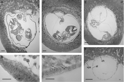
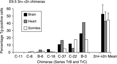
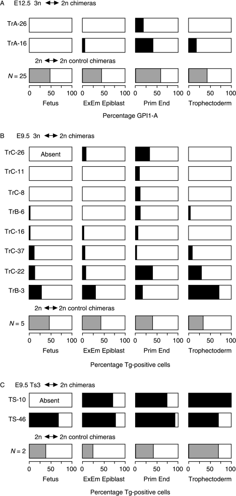
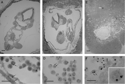
Human confined placental mosaicism (CPM), where the placental trophoblast is mosaic for a chromosome abnormality but the fetus is chromosomally normal, can cause problems for prenatal diagnosis, but its causes are poorly understood. Tetraploid<-->diploid chimeras provide a model for the development of one type of CPM, but animal models for other types of restricted mosaicism are needed. The objective of the present study was to evaluate triploid<-->diploid and trisomy-3<-->diploid chimeric mouse conceptuses as new models for investigating the development of restricted mosaicism. Novel stocks of mice were generated to produce triploid and trisomy-3 embryos that could be identified by DNA in situ hybridisation to a chromosome 3 transgenic marker. Triploid<-->diploid and trisomy-3<-->diploid mouse chimeras were produced by embryo aggregation, and the contribution of triploid or trisomy-3 cells was analysed in the fetus and extraembryonic tissues. Only two trisomy-3<-->diploid chimeras were analysed but trisomy-3 cells contributed well to all lineages, so these chimeras did not show restricted mosaicism. In contrast, triploid cells usually contributed poorly to all lineages in the ten 3n<-->2n chimeras analysed. They contributed more to the primitive endoderm derivatives than other lineages and were present in the primitive endoderm derivatives of all ten chimeras, but excluded from fetuses and trophectoderm derivatives in some cases. This pattern of restricted mosaicism differs from that reported for tetraploid cells in tetraploid<-->diploid chimeras, and triploid<-->diploid chimeras may provide a useful model for the development of some types of restricted mosaicism in human conceptuses.
Figures
References
-
- Azuma S, Fukuda Y, Toyoda Y. Studies on the production of 2n↔3n chimeric mouse embryos. II. Post-implantation development. Japanese Journal of Animal Reproduction. 1991;37:79–87.
-
- Bennett P, Vaughan J, Henderson D, Loughna S, Moore G. Association between confined placental trisomy, fetal uniparental disomy and early uterine growth retardation. Lancet. 1992;340:1284–1285. - PubMed
-
- Cattanach B, Moseley H. Nondisjunction and reduced fertility caused by tobacco mouse metacentric chromosomes. Cytogenetics and Cell Genetics. 1973;12:264–287. - PubMed
-
- Copp AJ. Death before birth. Clues from gene knockouts and mutations. Trends in Genetics. 1995;11:87–93. - PubMed
-
- Cox DR, Smith SA, Epstein LB, Epstein CJ. Mouse trisomy 16 as an animal model for human trisomy 21 (Down syndrome): production of viable trisomy 16↔diploid mouse embryos. Developmental Biology. 1984;101:416–424. - PubMed
Publication types
MeSH terms
Grants and funding
LinkOut - more resources
Full Text Sources
