The hypothetical protein Atu4866 from Agrobacterium tumefaciens adopts a streptavidin-like fold
- PMID: 18042676
- PMCID: PMC2144585
- DOI: 10.1110/ps.073167408
The hypothetical protein Atu4866 from Agrobacterium tumefaciens adopts a streptavidin-like fold
Abstract
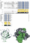
Atu4866 is a 79-residue conserved hypothetical protein of unknown function from Agrobacterium tumefaciens. Protein sequence alignments show that it shares > or =60% sequence identity with 20 other hypothetical proteins of bacterial origin. However, the structures and functions of these proteins remain unknown so far. To gain insight into the function of this family of proteins, we have determined the structure of Atu4866 as a target of a structural genomics project using solution NMR spectroscopy. Our results reveal that Atu4866 adopts a streptavidin-like fold featuring a beta-barrel/sandwich formed by eight antiparallel beta-strands. Further structural analysis identified a continuous patch of conserved residues on the surface of Atu4866 that may constitute a potential ligand-binding site.
Figures


References
-
- Bompard-Gilles, G., Remaut, H., Villeret, V., Prange, T., Fanuel, L., Delmarcelle, M., Joris, B., Frere, J.M., Van Beeumen, J. Crystal structure of a D-aminopeptidase from Ochrobactrum anthropi, a new member of the “penicillin-recognizing enzyme” family. Structure. 2000;8:971–980. - PubMed
-
- Brunger, A.T., Adams, P.D., Clore, G.M., DeLano, W.L., Gros, P., Grosse-Kunstleve, R.W., Jiang, L.S., Kuszewski, J., Nilges, M., Pannu, N.S., et al. Crystallography & NMR system: A new software suite for macromolecular structure determination. Acta Crystallogr. 1998;D54:905–921. - PubMed
-
- Cornilescu, G., Delaglio, F., Bax, A. Protein backbone angle restraints from searching a database for chemical shift and sequence homology. J. Biomol. NMR. 1999;13:289–302. - PubMed
-
- Delaglio, F., Grzesiek, S., Vuister, G.W., Zhu, G., Pfeifer, J., Bax, A. NMRPipe: A multidimensional spectral processing system based on UNIX pipes. J. Biomol. NMR. 1995;6:277–293. - PubMed
Publication types
MeSH terms
Substances
LinkOut - more resources
Full Text Sources
