. 2008 Jan;17(1):66-71.
doi: 10.1110/ps.073130208. Epub 2007 Nov 27.

Characterization of an exosite binding inhibitor of matrix metalloproteinase 13

Affiliations

Characterization of an exosite binding inhibitor of matrix metalloproteinase 13

Lata T Gooljarsingh et al. Protein Sci. 2008 Jan.

Abstract

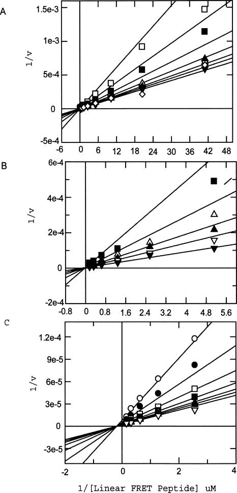
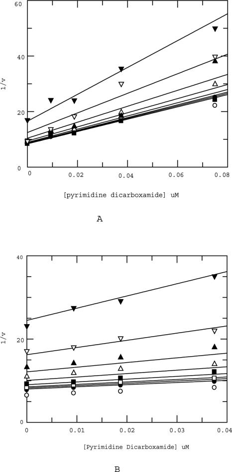
Matrix metalloproteinase 13 (MMP13) is a key enzyme implicated in the degradation of the extracellular matrix in osteoarthritis. Clinical administration of broad spectrum MMP inhibitors such as marimastat has been implicated in severe musculo-skeletal side effects. Consequently, research has been focused on designing inhibitors that selectively inhibit MMP13, thereby circumventing musculo-skeletal toxicities. A series of pyrimidine dicarboxamides were recently shown to be highly selective inhibitors of MMP13 with a novel binding mode. We have applied a molecular ruler to this exosite by dual inhibition studies involving a potent dicarboxamide in the presence of two metal chelators of different sizes. A larger hydroxamate mimic overlaps and antagonizes binding of the dicarboxamide to the exosite whereas the much smaller acetohydroxamate synergizes with the dicarboxamide. These studies elucidate the steric requirement for compounds that fit exclusively into the active site, a mandate for generating highly selective MMP13 inhibitors.


Figures

Figure 1.
Figure 1.
Structure of all three inhibitors discussed. (A) Pyrimidine dicarboxamide. (B) Hydroxamate mimic. (C) Acetohydroxamate.
Figure 2.
Figure 2.
(A) Overlaid structure of the catalytic domain of MMP13 with pyrimidine dicarboxamide (green) and hydroxamate mimic (magenta). The teal and gold ribbon diagrams represent MMP13 and exhibit a high degree of flexibility in the “selectivity loop” (denoted by *), which may be attributable to the presence of a glycine residue. The two inhibitors overlap by 6 Å. (B) Docked structure of MMP 13 with pyrimidine dicarboxamide (green) and acetohydroxamate (orange). These two docked structures are 6 Å apart.
Figure 3.
Figure 3.
(A) Acetohydroxamate fits best to a competitive model described in Materials and Methods. The same fit was also obtained (data not shown) when the THP substrate was used in lieu of the linear substrate. Legend (varying concentrations of inhibitor): □ 20 mM, ■ 10 mM, △ 5 mM, ▲ 2.5 mM, ◇ 1.25 mM, ▼ 0 mM. (B) Hydroxamate mimic fit best to a competitive model described in Materials and Methods. The same fit was also obtained (data not shown) when the THP substrate was used in lieu of the linear substrate. Legend (varying concentrations of inhibitor): ■ 2.5 nM, △ 1.25 nM, ▲ 0.625 nM, ▽ 0.3125 nM, ▼ 0 nM. (C) Pyrimdine dicarboxamide fit best to a noncompetitive model described in Materials and Methods. The same fit was also obtained (data not shown) when the THP substrate was used in lieu of the linear substrate. Legend (varying concentrations of inhibitor): ○ 125 nM, ● 62.5 nM, □ 31.25 nM, ■ 15.625 nM, △ 7.8125 nM, ▲ 3.90625 nM, ▽ 0 nM.
Figure 4.
Figure 4.
(A) Yonetani-Theorell analysis of pyrimidine dicarboxamide in the presence of hydroxamate mimic (linear substrate). Legend (varying concentrations of hydroxamate mimic): ○ 0 nM, ● 0.125 nM, □ 0.25 nM, ■ 0.5 nM, △ 0.75 nM, ▲ 1.25 nM, ▽ 2.5 nM. The B value from the fit was >>>1, indicating antagonism of binding of pyrimidine dicarboxamide by the hydroxamate mimic. (B) Yonetani-Theorell analysis of pyrimidine dicarboxamide in the presence of hydroxamate mimic (THP substrate). Legend (varying concentrations of hydroxamate mimic): ○ 0 nM, □ 0.25 nM, ■ 0.5 nM, △ 0.75 nM, ▲ 1.25 nM, ▽ 2.5 nM, ▼ 5 nM. The B value from the fit was >>>1, indicating antagonism of binding of pyrimidine dicarboxamide by the hydroxamate mimic.
Figure 5.
Figure 5.
(A) Yonetani-Theorell analysis of pyrimidine dicarboxamide in the presence of acetohydroxamate (linear substrate). Legend (varying concentrations of acetohydroxamate): ○ 0 μM, ● 156.25 μM, □ 312.5 μM, ■ 625 μM, △ 1250 μM, ▲ 2500 μM, ▽ 5000 μM, ▼ 10,000 μM. The B value from the fit was <1, indicating synergism of binding of pyrimidine dicarboxamide by the acetohydroxamate. (B) Yonetani-Theorell analysis of pyrimidine dicarboxamide in the presence of acetohydroxamate (THP substrate). Legend (varying concentrations of acetohydroxamate): ○ 0 μM, ● 156.25 μM, □ 312.5 μM, ■ 625 μM, △ 1250 μM, ▲ 2500 μM, ▽ 5000 μM, ▼ 10,000 μM. The B value from the fit was <1, indicating synergism of binding of pyrimidine dicarboxamide by the acetohydroxamate.

References

    1. Bigg, H.F., Rowan, A.D. The inhibition of metalloproteinases as a therapeutic target in rheumatoid arthritis and osteoarthritis. Curr. Opin. Pharmacol. 2001;1:314–320. - PubMed
    1. Brown, P.D. Ongoing trials with matrix metalloproteinase inhibitors. Expert Opin. Investig. Drugs. 2000;9:2167–2177. - PubMed
    1. Chung, L., Dinakarpandian, D., Yoshida, N., Lauer-Fields, J.L., Fields, G.B., Visse, R., Nagase, H. Collagenase unwinds triple-helical collagen prior to peptide bond hydrolysis. EMBO J. 2004;23:3020–3030. - PMC - PubMed
    1. Engel, C.K., Pirard, B., Schimanski, S., Kirsch, R., Habermann, J., Klinger, O., Schlotte, V., Weithmann, K.U., Wendt, K.U. Structural basis for the highly selective inhibition of MMP13. Chem. Biol. 2005;12:181–189. - PubMed
    1. Freskos, J.N., McDonald, J.J., Mischke, B.V., Mullins, P.B., Shieh, H.S., Stegeman, R.A., Stevens, A.M. Synthesis and identification of conformationally constrained selective MMP inhibitors. Bioorg. Med. Chem. Lett. 1999;9:1757–1760. - PubMed

Publication types

MeSH terms

Substances

LinkOut - more resources