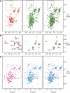
Solution NMR studies of apo-mSin3A and -mSin3B reveal that the PAH1 and PAH2 domains are structurally independent
- PMID: 18042683
- PMCID: PMC2144601
- DOI: 10.1110/ps.073097308
Solution NMR studies of apo-mSin3A and -mSin3B reveal that the PAH1 and PAH2 domains are structurally independent
Abstract
The evolutionarily conserved mammalian Sin3 (mSin3) transcriptional corepressor interacts with a diverse array of transcription factors mainly through two PAH (paired amphipathic helix) domains located near the N terminus. Previous studies suggested the possibility of interdomain interactions involving the PAH domains. Here, we show that the domains are structurally independent and the properties of the individual domains, such as the conformational heterogeneity and the ability of mSin3A PAH2 to homodimerize, are preserved in constructs that span both PAH domains. Our results thus suggest that the N-terminal segments of the Sin3 proteins are broadly available for interactions with other proteins and that the PAH domains are organized into structurally independent modules. Our data also rule out any heterotypic association between the paralogous mSin3A and mSin3B proteins via interactions involving the mSin3A PAH2 domain.
Figures

References
-
- Ayer, D.E. Histone deacetylases: Transcriptional repression with SINers and NuRDs. Trends Cell Biol. 1999;9:193–198. - PubMed
-
- Brubaker, K., Cowley, S.M., Huang, K., Loo, L., Yochum, G.S., Ayer, D.E., Eisenman, R.N., Radhakrishnan, I. Solution structure of the interacting domains of the Mad–Sin3 complex: Implications for recruitment of a chromatin-modifying complex. Cell. 2000;103:655–665. - PubMed
-
- Elgin, S.C.R., Workman, J.L. Chromatin structure and gene expression. 2d ed. Oxford University Press; New York: 2000. p. 328.
Publication types
MeSH terms
Substances
Grants and funding
LinkOut - more resources
Full Text Sources
