. 2007 Dec 4;104(49):19315-20.
doi: 10.1073/pnas.0709856104. Epub 2007 Nov 27.

Cryo-EM study of the spinach chloroplast ribosome reveals the structural and functional roles of plastid-specific ribosomal proteins

Affiliations

Cryo-EM study of the spinach chloroplast ribosome reveals the structural and functional roles of plastid-specific ribosomal proteins

Manjuli R Sharma et al. Proc Natl Acad Sci U S A. .

Abstract

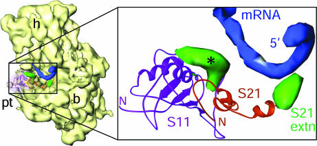
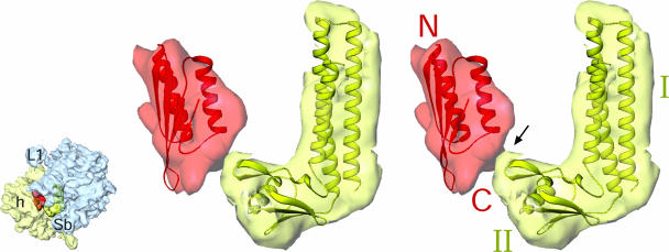
Protein synthesis in the chloroplast is carried out by chloroplast ribosomes (chloro-ribosome) and regulated in a light-dependent manner. Chloroplast or plastid ribosomal proteins (PRPs) generally are larger than their bacterial counterparts, and chloro-ribosomes contain additional plastid-specific ribosomal proteins (PSRPs); however, it is unclear to what extent these proteins play structural or regulatory roles during translation. We have obtained a three-dimensional cryo-EM map of the spinach 70S chloro-ribosome, revealing the overall structural organization to be similar to bacterial ribosomes. Fitting of the conserved portions of the x-ray crystallographic structure of the bacterial 70S ribosome into our cryo-EM map of the chloro-ribosome reveals the positions of PRP extensions and the locations of the PSRPs. Surprisingly, PSRP1 binds in the decoding region of the small (30S) ribosomal subunit, in a manner that would preclude the binding of messenger and transfer RNAs to the ribosome, suggesting that PSRP1 is a translation factor rather than a ribosomal protein. PSRP2 and PSRP3 appear to structurally compensate for missing segments of the 16S rRNA within the 30S subunit, whereas PSRP4 occupies a position buried within the head of the 30S subunit. One of the two PSRPs in the large (50S) ribosomal subunit lies near the tRNA exit site. Furthermore, we find a mass of density corresponding to chloro-ribosome recycling factor; domain II of this factor appears to interact with the flexible C-terminal domain of PSRP1. Our study provides evolutionary insights into the structural and functional roles that the PSRPs play during protein synthesis in chloroplasts.


Conflict of interest statement

The authors declare no conflict of interest.

Figures

Fig. 1.
Fig. 1.
Comparison of the cryo-EM maps of the spinach chloroplast 70S ribosome at 9.4-Å resolution and the E. coli 70S ribosome. (A and B) The chloro-ribosome with 30S subunit (yellow) and 50S subunit (green). (C and D) The E. coli 70S ribosome (8) with 30S subunit (pale yellow) and 50S subunit (blue). In A/C and B/D, ribosomes are shown from the L7/L12 and L1 sides, respectively. Structural differences of the 30S and 50S subunits are marked by red arrows. Landmarks of the 30S subunit: h, head; pt, platform; sh, shoulder; and sp, spur. Landmarks of the 50S subunit: CP, central protuberance; H38, 23S rRNA helix 38; L1, protein L1 protuberance; and Sb, stalk base.
Fig. 2.
Fig. 2.
Distribution of RNA and protein on the chloro-ribosome with positions of PSRPs and PRP extensions highlighted. (A and B) 30S subunit: pale yellow, rRNA; yellow, PRPs; green, PRP extensions; and red, PSRPs. (C and D) 50S subunit: blue, rRNA; purple, PRPs, green, PRP extensions; and red, PSRP5. In A/C and B/D, subunits are shown from the interface and solvent sides, respectively. To avoid visual complexity, only densities corresponding to PRP extensions (with suffix “ext”), conformational changes (with suffix “c”), or shifts in the position of PRPs (with suffix “s”) with respect to their bacterial orthologs, and PSRPs (–5), are labeled. Numbers following S or L identify small and large subunit PRPs, respectively, and numbers following h or H identify 16S and 23S rRNA helices within small and large subunits, respectively. Landmarks: b, body of the 30S subunit; pRRF, domain I of pRRF; St, L7/L12 stalk of the 50S subunit. All other landmarks are the same as in Fig. 1.
Fig. 3.
Fig. 3.
The mRNA exit channel. X-ray crystallographic structure of the whole 30S mRNA complex (19) was docked into the cryo-EM map of the chloro-30S subunit. Boxed region in the thumbnail (Left) is enlarged to show the 5′ terminus of mRNA (blue), PRPs S11 (magenta ribbons), and S21 (orange ribbons) with their extensions (green). The CTE of S21 is close enough to make direct contact with the mRNA, whereas the extra density associated with S11 (marked with an asterisk) is within 13 Å of the mRNA. The latter density could be partly attributable to a local conformational change.
Fig. 4.
Fig. 4.
Stereoview presentation of the interaction between CTD of PSRP1 with domain II of pRRF. Difference map, showing the N-terminal domain of PSRP1 (semitransparent red), is shown with the fitted homology model (red ribbons). N-terminal (N) and C-terminal (C) ends are labeled. Difference map corresponding to pRRF (semitransparent yellow) is shown with fitted atomic structure (greenish-yellow ribbons) of Thermotoga maritima RRF (PDB ID 1DD5). Two domains (I and II) of RRF are labeled. Thumbnail to the left depicts the orientation of the chloro-ribosome with small (semitransparent pale yellow) and large (semitransparent blue) subunits identified.
Fig. 5.
Fig. 5.
Topography of the polypeptide-exit tunnel in the 50S subunit. The modeled polypeptide chain (magenta) exiting from the tunnel is shown. PRPs surrounding the tunnel exit are shown as semitransparent purple masses with homology models (shown as ribbons) of PRPs docked in. Light blue density corresponds to 23S rRNA. Thumbnail to the left shows the orientation of the ribosome with the boxed region, which has been enlarged for stereoviewing.

References

    1. Harris EH, Boynton JE, Gillham NW. Microbiol Rev. 1994;58:700–754. - PMC - PubMed
    1. O'Brien TW. Gene. 2002;286:73–79. - PubMed
    1. Schmitz-Linneweber C, Maier RM, Alcaraz JP, Cottet A, Herrmann RG, Mache R. Plant Mol Biol. 2001;45:307–315. - PubMed
    1. Margulis L. Origin of Eukaryotic Cells. New Haven: Yale Univ Press; 1970.
    1. Yamaguchi K, Subramanian AR. J Biol Chem. 2000;275:28466–28482. - PubMed

Publication types

MeSH terms