. 2007 Dec 18;104(51):20454-9.
doi: 10.1073/pnas.0708102104. Epub 2007 Nov 27.

A dual function for a bacterial small RNA: SgrS performs base pairing-dependent regulation and encodes a functional polypeptide

Affiliations

A dual function for a bacterial small RNA: SgrS performs base pairing-dependent regulation and encodes a functional polypeptide

Caryn S Wadler et al. Proc Natl Acad Sci U S A. .

Abstract

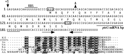
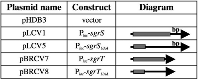
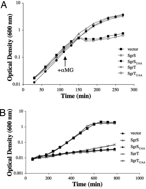
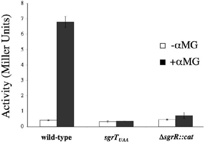
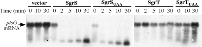
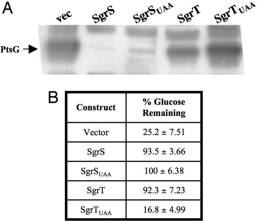
SgrS is a 227-nt small RNA that is expressed in Escherichia coli during glucose-phosphate stress, a condition associated with intracellular accumulation of glucose-6-phosphate caused by disruption of glycolytic flux. Under stress conditions, SgrS negatively regulates translation and stability of the ptsG mRNA, encoding the major glucose transporter, by means of a base pairing-dependent mechanism requiring the RNA chaperone Hfq. SgrS activity mitigates the effects of glucose-phosphate stress, and the present study has elucidated a function of SgrS that is proposed to contribute to the stress response. The 5' end of SgrS, upstream of the nucleotides involved in base pairing with the ptsG mRNA, contains a 43-aa ORF, sgrT, that is conserved in most species that contain SgrS-like small RNAs. The sgrT gene is translated in E. coli under conditions of glucose-phosphate stress. Analysis of alleles that separate the base pairing function of SgrS from the sgrT coding sequence revealed that either of these functions alone are sufficient for previously characterized SgrS phenotypes. SgrS-dependent down-regulation of ptsG mRNA stability does not require SgrT and SgrT by itself has no effect on ptsG mRNA stability. Cells expressing sgrT alone had a defect in glucose uptake even though they had nearly wild-type levels of PtsG (IICB(Glc)). Together, these data suggest that SgrS represents a previously unrecognized paradigm for small RNA (sRNA) regulators as a bifunctional RNA that encodes physiologically redundant but mechanistically distinct functions contributing to the same stress response.


Conflict of interest statement

The authors declare no conflict of interest.

Figures

Fig. 1.
Fig. 1.
The SgrS sRNA contains a conserved ORF, sgrT. (Upper) The SgrS sequence is shown with the translated product of sgrT below the nucleotide sequence. The putative ribosome binding site for sgrT is indicated by the horizontal bar labeled “RBS.” The start and stop codons for sgrT are boxed. The fifth codon of sgrT was mutated to a “TAA” by a single base pair substitution indicated by the arrow and “A” (described in the text and in Fig. 2). The sequences involved in base pairing with the ptsG mRNA (7) are indicated by the horizontal line (labeled “ptsG mRNA bp”) below the nucleotide sequences. The inverted repeat that forms the terminator at the 3′ end of SgrS is indicated by horizontal arrows below the nucleotide sequence. (Lower) The SgrT amino acid sequences from E. coli K12 (K12) and other bacterial species were aligned with ClustalW. Matches to the consensus are shaded. CFT073, E. coli CFT073; S.e., Salmonella enterica paratyphi ATCC9150; S.ty., Salmonella enterica serovar Typhi Ty2; K.p., Klebsiella pneumoniae KP32; E.ca., Erwinia carotovora atroseptica SCRI1043; A.h., Aeromonas hydrophila ATCC7966.
Fig. 2.
Fig. 2.
Construction of alleles that separate base pairing and SgrT functions. Plasmids are described in more detail in Methods and SI Table 1. All constructs are under the control of the Plac promoter. The SgrS molecule is depicted as an arrow, where the arrowhead is the 3′ end of SgrS. The location of sgrT is represented by the shaded rectangle at the 5′ end, and the location of the base pairing sequences at the 3′ end is indicated by the label “bp.” sgrSUAA contains a stop codon that truncates the sgrT ORF. The sgrT construct lacks the base pairing region. The sgrTUAA construct lacks the base pairing region and also contains a truncated sgrT ORF.
Fig. 3.
Fig. 3.
Effect of SgrS base pairing and SgrT on recovery from glucose-phosphate stress and growth in glucose minimal medium. (A) A ΔsgrS::kan, lacIq+ host strain (CV104) carrying plasmids (pHDB3, pLCV1, pLCV5, pBRCV7, and pBRCV8) with alleles described in Fig. 2 was grown in LB with ampicillin and IPTG. Cells were stressed by addition of 0.5% αMG at early log phase. The data shown are representative of at least three independent experiments. (B) The strains described in A were grown in minimal A medium with glucose in the presence of ampicillin and IPTG. The results shown are representative of at least three independent experiments.
Fig. 4.
Fig. 4.
Translation of sgrT is activated under stress conditions in an SgrR-dependent manner. All strains carry a translational sgrT′-′lacZ fusion at the native sgrT locus. Strains BH300 (wild-type), BH301 (sgrTUAA), and BH302 (ΔsgrR::cat) were grown in rich medium to mid-log phase and split, and half of each culture was exposed to 0.005% αMG. β-galactosidase activity was measured at several time points thereafter. The data displayed are the average of three experimental trials at 3 h after exposure.
Fig. 5.
Fig. 5.
SgrT does not affect levels of ptsG mRNA. The ΔsgrS::kan, lacIq+ strain CV104 carrying constructs depicted in Fig. 2 was grown to mid-log phase in LB with ampicillin. Northern blot analysis was performed on total RNA extracts harvested at times indicated after cells were exposed to IPTG to induce expression of sgrS and sgrT constructs. The blot was probed for ptsG mRNA (indicated at the left).
Fig. 6.
Fig. 6.
SgrS base pairing and SgrT functions individually block glucose uptake by different mechanisms. (A) Strains carrying plasmid constructs (as in Fig. 2) were grown in MOPS defined medium with glucose and amino acids with IPTG. Western blot analysis was performed on total protein extracts from samples harvested after 5 h of growth. The blot was probed for PtsG by using an αIIBGlc antibody. The position of the PtsG (IICBGlc) protein is indicated at left. Bands above and below the PtsG band are cross-reacting proteins and served as loading controls. (B) Strains are as described in A. Cells were harvested after 5 h of growth, and the amount of glucose remaining in the medium was measured. The numbers reported represent the amount of glucose remaining at 5 h divided by the amount of glucose in media before inoculation (% glucose remaining). The average of three independent experiments is reported.

Comment in

  • Small RNAs making a small protein.
    Morita T, Aiba H. Morita T, et al. Proc Natl Acad Sci U S A. 2007 Dec 18;104(51):20149-50. doi: 10.1073/pnas.0710634105. Epub 2007 Dec 11. Proc Natl Acad Sci U S A. 2007. PMID: 18077354 Free PMC article. No abstract available.

References

    1. Englesberg E, Anderson RL, Weinberg R, Lee N, Hoffee P, Huttenhauer G, Boyer H. J Bacteriol. 1962;84:137–146. - PMC - PubMed
    1. Kadner RJ, Murphy GP, Stephens CM. J Gen Microbiol. 1992;138:2007–2014. - PubMed
    1. Yarmolinsky MB, Wiesmeyer H, Kalckar HM, Jordon E. Proc Natl Acad Sci USA. 1959;45:1786–1791. - PMC - PubMed
    1. Kimata K, Tanaka Y, Inada T, Aiba H. EMBO J. 2001;20:3587–3595. - PMC - PubMed
    1. Morita T, El-Kazzaz W, Tanaka Y, Inada T, Aiba H. J Biol Chem. 2003;278:15608–15614. - PubMed

MeSH terms