. 2007 Dec 4;104(49):19613-8.
doi: 10.1073/pnas.0705147104. Epub 2007 Nov 27.

CERK1, a LysM receptor kinase, is essential for chitin elicitor signaling in Arabidopsis

Affiliations

CERK1, a LysM receptor kinase, is essential for chitin elicitor signaling in Arabidopsis

Ayako Miya et al. Proc Natl Acad Sci U S A. .

Abstract

Chitin is a major component of fungal cell walls and serves as a microbe-associated molecular pattern (MAMP) for the detection of various potential pathogens in innate immune systems of both plants and animals. We recently showed that chitin elicitor-binding protein (CEBiP), plasma membrane glycoprotein with LysM motifs, functions as a cell surface receptor for chitin elicitor in rice. The predicted structure of CEBiP does not contain any intracellular domains, suggesting that an additional component(s) is required for signaling through the plasma membrane into the cytoplasm. Here, we identified a receptor-like kinase, designated CERK1, which is essential for chitin elicitor signaling in Arabidopsis. The KO mutants for CERK1 completely lost the ability to respond to the chitin elicitor, including MAPK activation, reactive oxygen species generation, and gene expression. Disease resistance of the KO mutant against an incompatible fungus, Alternaria brassicicola, was partly impaired. Complementation with the WT CERK1 gene showed cerk1 mutations were responsible for the mutant phenotypes. CERK1 is a plasma membrane protein containing three LysM motifs in the extracellular domain and an intracellular Ser/Thr kinase domain with autophosphorylation/myelin basic protein kinase activity, suggesting that CERK1 plays a critical role in fungal MAMP perception in plants.


Conflict of interest statement

The authors declare no conflict of interest.

Figures

Fig. 1.
Fig. 1.
Characterization of CERK1 and its mutants. (A) Predicted structure of CERK1 protein and the gene model of At3g21630 including the positions of T-DNA and Ds-transposon insertion. LysM, LysM motif; TM, transmembrane domain; JM, juxtamembrane domain. (B) Expression of CERK1 in the Ds (cerk1-1, Left) and T-DNA (cerk1-2, Right) insertional mutants. Primers for RT-PCR were designed to amplify the region containing the positions for Ds or T-DNA insertion. (C) Induction of the expression of CERK1 gene by chitin elicitor treatment. The Arabidopsis seedlings were treated with 100 μg·ml−1 of (GlcNAc)8, taken at the indicated time intervals and used for RT-PCR as described in the text.
Fig. 2.
Fig. 2.
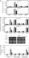
Specific loss of chitin-induced ROS generation in cerk1 mutants and the recovery in the transformants complemented with the WT CERK1 gene. (A) ROS generation by cerk1-1 and cerk1-2 mutants treated with 100 μg·ml−1 of (GlcNAc)8. ROS was analyzed at 0, 30, 60, and 120 min after the elicitor treatment (shown in white, stripe, gray, and black, respectively). WT, WT A. thaliana; GN8, N-acethylchitooctaose, DW, control seedlings treated with water. (B) ROS generation by cerk1-1 and cerk1-2 mutants treated with 100 μg·ml−1 of P. aeruginosa LPS. (C) Recovery of CERK1 expression in the cerk1-1 mutants complemented with WT CERK1 gene. (D) Recovery of chitin-induced ROS generation in the same transformants.
Fig. 3.
Fig. 3.
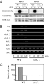
Loss of chitin elicitor responses in cerk1-1 mutant. (A) Loss of MPK3 and MPK6 activation by (GlcNAc)8 in cerk1-1 mutant. (Top) Immunocomplex kinase assay for monitoring MPK3 and MPK6 activities upon (GlcNAc)8 elicitation. Seedlings (0.1 g) of WT and cerk1-1 grown in MGRL media for 8 days were transferred on the same media in plastic plates and then incubated for 24 h at 22°C to eliminate mechanical shock. The seedlings were treated with either 100 μg·ml−1 of (GlcNAc)8 (+) or MGRL media as a mock (−) for 10 min, then frozen in liquid nitrogen. MAPK activity was examined by immunocomplex kinase assay as described in the experimental procedures. (Middle) Western blot analysis of MPK3 and MPK6 proteins in WT and cerk1-1. The same protein extracts (20 μg) for the immunocomplex kinase assay were used for Western blot analysis. Equal protein loading was confirmed by staining membrane with the Coomassie brilliant blue R-250 (Lower). (B) Defense gene expression induced by chitin elicitor treatment in WT and cerk1-1 seedlings. Seedlings were treated with 100 μg·ml−1 of (GlcNAc)8 and taken at indicated time intervals for RT-PCR analysis. (C) Analysis of global gene expression induced by chitin elicitor treatment in WT and cerk1-1 seedlings. Seedlings were treated with 100 μg·ml−1 of (GlcNAc)8 for 2 h and used for RNA extraction. Only three genes in cerk1-1 mutant showed the significant up-regulation by the elicitor treatment compared with 1,222 genes in WT. Similarly, only two genes showed down-regulation in cerk1-1, compared with 421 genes in WT.
Fig. 4.
Fig. 4.
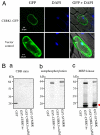
CERK1 encodes a functional protein kinase localized at the plasma membrane. (A) Detection of CERK1-GFP fusion protein at the plasma membrane. The CERK1-GFP fusion protein was transiently expressed in onion epidermal cells by particle bombardment and observed with a confocal laser-scanning microscope. The vector control expressing GFP was similarly observed. (B) Protein kinase activity of intracellular region of CERK1. (a) Coomassie brilliant blue (CBB) staining of purified samples. The GST alone, GST-CERK1Int, GST- CERK1IntΔJM, and GST-SYMRK were expressed in E. coli and purified. The samples (0.5 μg) were separated by 4–12% gradient polyacrylamide gel and stained with CBB R-250. (b) Autophosphorylation activity of GST-CERK1Int. The samples (0.5 μg) were incubated in a reaction buffer containing [γ-32P] at 30°C for 30 min. The reaction was stopped by addition of the SDS sample buffer. Samples were subsequently separated by SDS/PAGE as described above and autoradiographed. (c) MBP-phosphorylation activity of GST-CERK1Int. The in vitro phosphorylation assay was performed as b but in the presence of MBP (5 μg).

References

    1. Heath MC. Curr Opin Plant Biol. 2000;3:315–319. - PubMed
    1. Nürnberger T, Brunner F, Kemmerling B, Piater L. Immunol Rev. 2004;198:249–266. - PubMed
    1. Chisholm ST, Coaker G, Day B, Staskawicz BJ. Cell. 2006;124:803–814. - PubMed
    1. Nürnberger T, Lipka V. Mol Plant Pathol. 2005;6:335–345. - PubMed
    1. Jones JD, Dangl JL. Nature. 2006;444:323–329. - PubMed

Publication types

MeSH terms

Associated data