. 2007 Dec 4;104(49):19238-43.
doi: 10.1073/pnas.0703854104. Epub 2007 Nov 27.

Epidermal growth factor receptor juxtamembrane region regulates allosteric tyrosine kinase activation

Affiliations

Epidermal growth factor receptor juxtamembrane region regulates allosteric tyrosine kinase activation

Kristina W Thiel et al. Proc Natl Acad Sci U S A. .

Abstract

Structural studies of the extracellular and tyrosine kinase domains of the epidermal growth factor receptor (ErbB-1) provide considerable insight into facets of the receptor activation mechanism, but the contributions of other regions of ErbB-1 have not been ascertained. This study demonstrates that the intracellular juxtamembrane (JM) region plays a vital role in the kinase activation mechanism. In the experiments described herein, the entire ErbB-1 intracellular domain (ICD) has been expressed in mammalian cells to explore the significance of the JM region in kinase activity. Deletion of the JM region (DeltaJM) results in a severe loss of ICD tyrosine phosphorylation, indicating that this region is required for maximal activity of the tyrosine kinase domain. Coexpression of DeltaJM and dimerization-deficient kinase domain ICD mutants revealed that the JM region is indispensable for allosteric kinase activation and productive monomer interactions within a dimer. Studies with the intact receptor confirmed the role of the JM region in kinase activation. Within the JM region, Thr-654 is a known protein kinase C (PKC) phosphorylation site that modulates kinase activity in the context of the intact ErbB-1 receptor; yet, the mechanism is not known. Whereas a T654A mutation promotes increased ICD tyrosine phosphorylation, the phosphomimetic T654D mutant generates a 50% reduction in ICD tyrosine phosphorylation. Similar to the DeltaJM mutants, the T654D mutant ICD failed to interact with a wild-type monomer. This study reveals an integral role for the intracellular JM region of ErbB-1 in allosteric kinase activation.


Conflict of interest statement

The authors declare no conflict of interest.

Figures
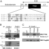
Fig. 1.
Fig. 1.
Tyrosine phosphorylation of ErbB-1 ICD. (A) Diagram of the ErbB receptors with alignment of JM regions. Basic residues are shaded gray; PKC phosphorylation sites in ErbB-1 (6, 7) and ErbB-2 (32) and a putative PKC phosphorylation site in ErbB-4 are boldface. TM, transmembrane domain; CT, C-terminal domain. (B) Lysates of Cos-7 cells transfected with ErbB-4 or ErbB-1 ICDs were precipitated (IP) with the indicated antibodies followed by Western blotting (WB) with a phosphotyrosine antibody (PY99; Upper). Blots were stripped and reprobed with the indicated antibodies to detect either ErbB-4 ICD or ErbB-1 ICD (Lower). Similar results were obtained when lysates were directly blotted with anti-PY99 (data not shown). (C) Tyrosine phosphorylation of PKC-site mutants. The indicated ErbB-1 ICD constructs were expressed as in B, precipitated with anti-ErbB-1, blotted with anti-PY99 (Upper), stripped, and reprobed with anti-FlagM2 (Lower). Relative pY, tyrosine phosphorylation (pY) relative to wild-type after normalizing for expression levels.
Fig. 2.
Fig. 2.
Tyrosine phosphorylation of JM deletion mutants. (A) JM residues in ErbB-1. Basic residues are shaded and Thr-654 is boldface as in Fig. 1. Arrows denote the N-terminal residue of the two JM deletions in the ICD, Δ645–662 and Δ645–676. The asterisk indicates the N-terminal residue of the TKD construct used by Zhang et al. (18) and Stamos et al. (20) for crystallography. (B) Indicated ICD constructs were transiently transfected into Cos-7 cells. Cell lysates were precipitated with anti-FlagM2 and blotted with anti-phosphotyrosine (PY99) or anti-FlagM2. (C) In vitro kinase assay comparing phosphorylation of ΔJM mutants with wild-type and K721R ICDs. After expression in Cos-7 cells, lysis, and precipitation with anti-FlagM2, the immunoprecipitates were incubated with [γ-32P]ATP (Upper) as described in Materials and Methods. Lysates were probed with anti-FlagM2 to confirm equal expression (Lower).
Fig. 3.
Fig. 3.
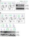
Ability of JM mutants to act as acceptor or donor monomers. (A Left) Cartoon of ICD mutants and predicted associations in coexpression experiments. Corresponding data lanes (A Right) are listed in brackets. Circles, V924R C-lobe mutation; stars, I682Q N-lobe mutation; ×, K721R mutation; green text, acceptor monomer; blue text, donor monomer. (A Right) Acceptor (V924R) and donor (I682Q) monomers were expressed alone (lanes 3 and 4) or coexpressed (lanes 5 and 6). Lysates were precipitated with anti-FlagM2 and blotted with either anti-PY99 or anti-FlagM2. (B Upper) Cartoon of potential interactions of donor (I682Q) and acceptor (V924R) monomers with ΔJM mutants containing additional N-lobe (I682Q) or C-lobe (V924R) mutations. (B Lower) ΔJM mutants having N-lobe or C-lobe mutations were coexpressed with acceptor (V924R) or donor (I682Q) monomers with intact JM regions.
Fig. 4.
Fig. 4.
Failure of ΔJM and T654D ICD mutants to interact with wild-type. (A) Flag-tagged wild-type and ΔJM mutant ICDs were transfected individually or cotransfected with wild-type Myc ICD as described in Materials and Methods. Lysates were precleared on protein G-Sepharose beads, precipitated with anti-Myc, and blotted with anti-FlagM2. The blot was stripped and reprobed with anti-EGFR. Lysates were also blotted with anti-EGFR. (B) T654D ICD-Flag was transfected with or without wild-type ICD-Myc as in A, and precleared lysates were precipitated with anti-FlagM2 followed by blotting with anti-Myc. Expression was assessed as in A.
Fig. 5.
Fig. 5.
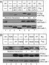
Role of JM region in EGF-stimulated allosteric kinase activation. (A) NIH 3T3 cells were transiently transfected with the indicated constructs. Forty hours later, cells were starved for 60 min in serum-free media and treated with EGF (50 ng/ml) for 5 min. Lysates were immediately immunoblotted with either phosphosite-specific ErbB-1 PY1173 (Upper) or ErbB-1 (Lower). (B and C) Experiments assessing the ability of JM deletions to act either as donors (B) or acceptors (C). The exposure time for C was longer than that in B to detect EGF-dependent tyrosine phosphorylation in lanes 5–8.

References

    1. Jae Song, Kil CC. J Cell Physiol. 2000;185:47–60. - PubMed
    1. Kil SJ, Hobert M, Carlin C. J Biol Chem. 1999;274:3141–3150. - PubMed
    1. He C, Hobert M, Friend L, Carlin C. J Biol Chem. 2002;277:38284–38293. - PubMed
    1. Lin S-Y, Makino K, Xia W, Matin A, Wen Y, Kwong KY, Bourguignon L, Hung M-C. Nat Cell Biol. 2001;3:802–808. - PubMed
    1. Martin-Nieto J, Villalobo A. Biochemistry. 1998;37:227–236. - PubMed

Publication types

MeSH terms