. 2008 Jan;63(1):26-32.
doi: 10.1203/PDR.0b013e31815b690d.

Role of matrix metalloproteinase-2 in newborn mouse lungs under hypoxic conditions

Affiliations

Role of matrix metalloproteinase-2 in newborn mouse lungs under hypoxic conditions

Namasivayam Ambalavanan et al. Pediatr Res. 2008 Jan.

Abstract

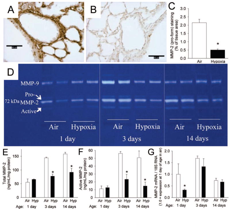
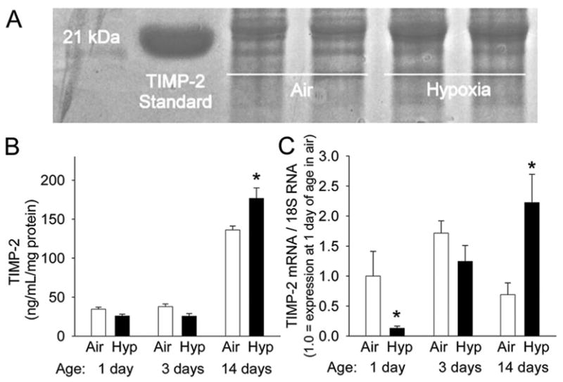
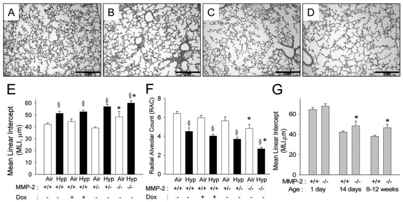
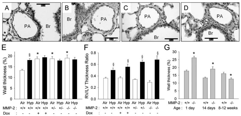
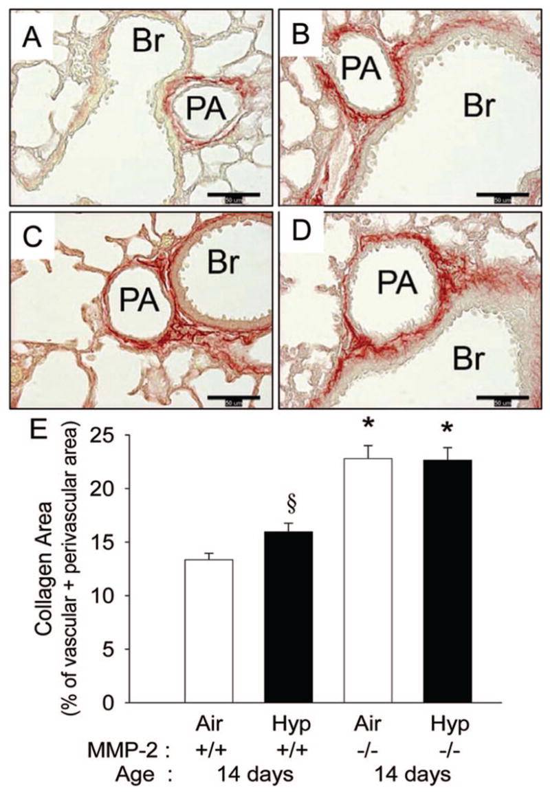
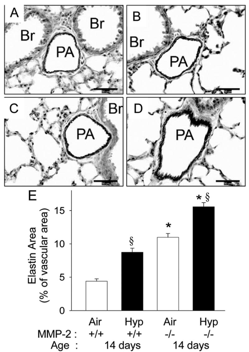
Hypoxia impairs normal neonatal pulmonary artery remodeling and alveolar development. Matrix metalloproteinase-2 (MMP-2), which regulates collagen breakdown, is important during development. Our objective was to test the hypothesis that hypoxia attenuates the normal postnatal increase in MMP-2 and evaluate alveolar development and pulmonary arterial remodeling in Mmp2 mice. C57BL/6 wild-type (WT), Mmp2, Mmp2, and MMP-inhibited (with doxycycline) mice were exposed to hypoxia (12% O2) or air from birth to 2 wk of age. Pulmonary arterial remodeling, alveolar development, and vascular collagen and elastin were evaluated. MMP-2 was estimated by quantitative real-time polymerase chain reaction, enzyme-linked immunosorbent assay, immunohistochemistry, and zymography. We observed that 1) in WT mice, hypoxia led to thicker-walled pulmonary arteries and impaired alveolarization, accompanied by decreased MMP-2 and increased tissue inhibitor of metalloproteinases-2 (TIMP-2); 2) Mmp2 mice in air had thicker-walled arteries, impaired alveolarization, and increased perivascular collagen and elastin compared with WT; 3) hypoxia further inhibited alveolarization but did not alter arterial thickening in Mmp2 mice. Mmp2 and MMP-inhibited mice also had thicker-walled arteries than WT in air, but alveolarization was not different. We conclude that hypoxia reduces the postnatal MMP-2 increase in the lung, which may contribute to abnormal pulmonary arterial remodeling and impaired alveolarization.


Figures

Figure 1
Figure 1
MMP-2 in the developing postnatal WT mouse lung and the effects of hypoxia (12% O2). (A, B) Immunohistochemical staining for proMMP-2 (brown) in lung sections from 14 d mice exposed to (A) air or (B) hypoxia (magnification, ×400; calibration bars = 50 μm). (C) Pro-MMP-2 staining quantitation as percent of tissue area (n = 6 mice/gp; mean ± SE; *p < 0.001). (D) Zymogram of lung extracts from mice exposed to air or hypoxia for 1, 3, and 14 d from birth. (E) Total MMP-2 measured by ELISA from lung extracts of mice exposed to air or hypoxia (Hyp) from 1 d for up to 14 d (n = 6 mice/gp; mean ± SE, *p < 0.05 vs air at same time point). (F) Active MMP-2 measured by ELISA from same samples as E (n = 6 mice/gp; mean ± SE, *p < 0.05 vs air at same time point). (G) Mmp2 mRNA by competitive real-time PCR in homogenized lungs from mice exposed to air or hypoxia from 1 d for up to 14 d (n = 6 mice/gp; mean ± SE, *p < 0.05 vs air at same time point).
Figure 2
Figure 2
TIMP-2 in the developing postnatal WT mouse lung and the effects of hypoxia. (A) Reverse zymogram for TIMPs in lung extracts from 14 d mice exposed to air or hypoxia from birth. (B) Densitometric quantitation of reverse zymograms for TIMP-2 from lung extracts of mice exposed to air or hypoxia (Hyp) from 1 d up to 14 d (n = 6 mice/gp; mean ± SE, *p < 0.05 vs air at same time point). (C) Timp2 mRNA by competitive real-time PCR in homogenized lungs from mice exposed to air or hypoxia from 1 d up to 14 d (n = 6 mice/gp; mean ± SE, *p < 0.05 vs air at same time point).
Figure 3
Figure 3
MMP-2 and alveolar development in WT and Mmp2−/− mice. (AD) Representative photomicrographs of mouse lungs from WT (A, B) and Mmp2−/− mice (C, D) after 14 d of air (A, C) or hypoxia (B, D) exposure (magnification, ×100; calibration bars = 250 μm). Alveoli are small and well developed with many secondary crests in WT air mice (A), large with fewer secondary crests in WT hypoxia mice (B) and Mmp2−/− air (C) mice, and are much larger with minimal septation in Mmp2−/− hypoxic mice (D). (E) MLI and (F) RAC at 14 d of age in WT (+/+) mice given either vehicle or doxycycline (Dox), Mmp2+/−, and Mmp2−/− mice after air and hypoxic (Hyp) exposure (mean ± SE; n = 6 mice/gp; *p < 0.05 vs corresponding WT controls, §p < 0.05 vs corresponding air). (G) MLI in WT and Mmp2−/− mice at 1 d, 14 d, and 8 –12 wk of age (mean ± SE; n = 6 mice/gp; *p < 0.05 vs WT at same time point).
Figure 4
Figure 4
MMP-2 and pulmonary arterial remodeling in WT and Mmp2−/− mice. (AD) Representative photomicrographs of mouse lung pulmonary arteries from WT (A, B) and Mmp2−/− mice (C, D) after 14 d of air (A, C) or hypoxic (B, D) exposure (magnification, ×400; calibration bars = 50 μm; PA, pulmonary artery; Br, bronchus). The wall thickness of the pulmonary artery is less in the WT air mice (A) than in WT hypoxia (B), Mmp2−/− air (C), or Mmp2−/− hypoxic mice (D). (E) Wall thickness (%) of pulmonary arteries and (F) right ventricle/left ventricle (RV/LV) thickness ratio at 14 d of age in WT mice given either vehicle or doxycycline (Dox), Mmp2+/−, and Mmp2−/− mice after air and hypoxic exposure (mean ± SE; n = 6 mice/gp; *p < 0.05 vs corresponding WT controls, §p < 0.05 vs corresponding air). (G) Wall thickness (%) in air exposed WT and Mmp2−/− mice at 1 d, 14 d, and 8 –12 wk of age (mean ± SE; n = 120 vessels/gp; *p < 0.05 vs WT at same time point).
Figure 5
Figure 5
Perivascular collagen in 14 d WT and Mmp2−/− mice. (AD) Representative photomicrographs of picric acid-Sirius red stained sections (collagen is stained red) of pulmonary arteries from WT (A, B) and Mmp2−/− mice (C, D) after 14 d of air (A, C) or hypoxic (B, D) exposure (magnification, ×400; calibration bars = 50 μm; PA, pulmonary artery; Br, bronchus). Collagen in the perivascular area is least in the WT air mice (A), increased in WT hypoxia (B), and further increased in Mmp2−/− air (C) and in Mmp2−/− hypoxic mice (D). (E) Collagen area measured by quantitative image analysis at 14 d of age in WT and Mmp2−/− mice after air and hypoxic exposure (mean ± SE; n = 6 mice/gp; *p < 0.05 vs corresponding WT controls, §p < 0.05 vs corresponding air).
Figure 6
Figure 6
Vascular elastin in 14 d WT and Mmp2−/− mice. (AD) Representative photomicrographs of elastic tissue stained sections (elastin is stained black) of pulmonary arteries from WT (A, B) and Mmp2−/− mice (C, D) after 14 d of air (A, C) or hypoxia (B, D) exposure (magnification, ×400; calibration bars = 50 μm; PA, pulmonary artery; Br, bronchus). Elastin is least in the WT air mice (A), increased in WT hypoxia (B), further increased in Mmp2−/− air (C), and is maximal in Mmp2−/− hypoxic mice (D). (E) Elastin area measured by quantitative image analysis at 14 d of age in WT and Mmp2−/− mice after air and hypoxic exposure (mean ± SE; n = 6 mice/gp; *p < 0.05 vs corresponding WT controls, §p < 0.05 vs corresponding air).

Similar articles

Cited by

References

    1. Haworth SG, Hislop AA. Lung development-the effects of chronic hypoxia. Semin Neonatol. 2003;8:1–8. - PubMed
    1. Walsh-Sukys MC. Persistent pulmonary hypertension of the newborn. The black box revisited. Clin Perinatol. 1993;20:127–143. - PubMed
    1. Goodman G, Perkin RM, Anas NG, Sperling DR, Hicks DA, Rowen M. Pulmonary hypertension in infants with bronchopulmonary dysplasia. J Pediatr. 1988;112:67–71. - PubMed
    1. Hoffmeister HM, Apitz J, Hoffmeister HE, Fischbach H. The correlation between blood pressure and morphometric findings in children with congenital heart disease and pulmonary hypertension. Basic Res Cardiol. 1981;76:647–656. - PubMed
    1. Steinhorn RH, Fineman JR. The pathophysiology of pulmonary hypertension in congenital heart disease. Artif Organs. 1999;23:970–974. - PubMed

Publication types

MeSH terms