. 2007 Nov 28;2(11):e1227.
doi: 10.1371/journal.pone.0001227.

Transforming growth factor: beta signaling is essential for limb regeneration in axolotls

Affiliations

Transforming growth factor: beta signaling is essential for limb regeneration in axolotls

Mathieu Lévesque et al. PLoS One. .

Abstract

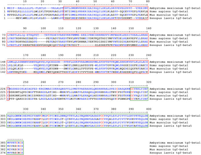
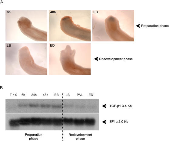
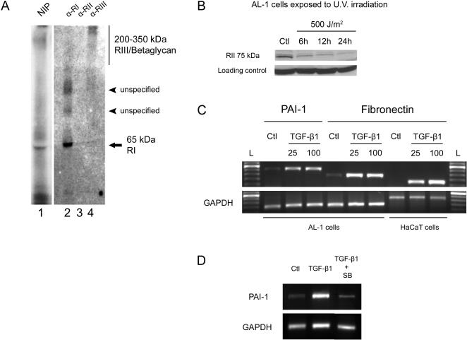
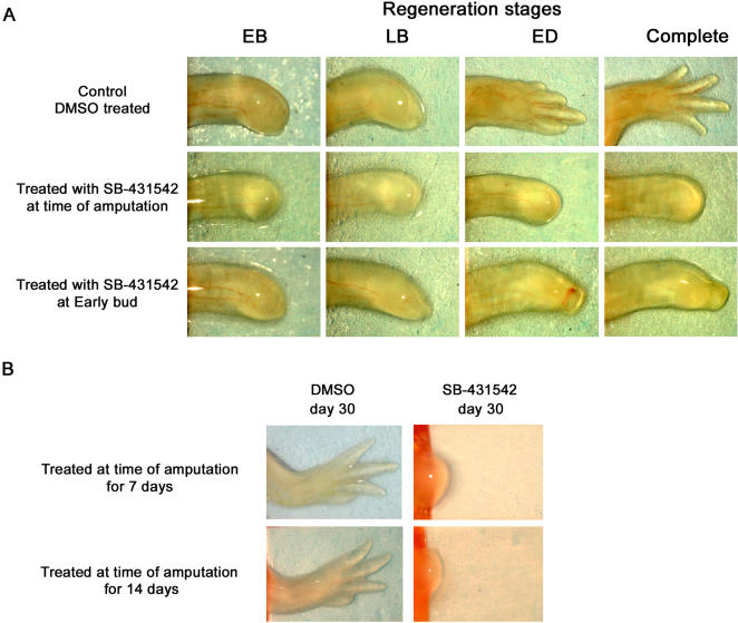
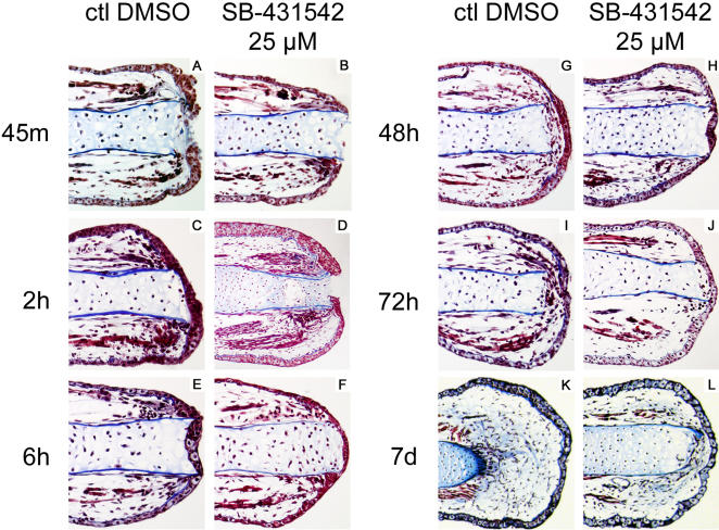
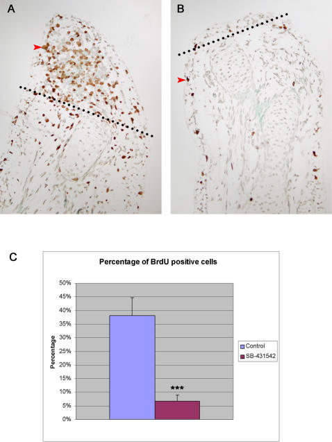
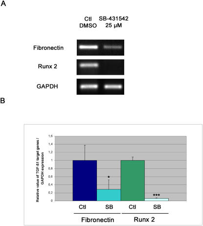
Axolotls (urodele amphibians) have the unique ability, among vertebrates, to perfectly regenerate many parts of their body including limbs, tail, jaw and spinal cord following injury or amputation. The axolotl limb is the most widely used structure as an experimental model to study tissue regeneration. The process is well characterized, requiring multiple cellular and molecular mechanisms. The preparation phase represents the first part of the regeneration process which includes wound healing, cellular migration, dedifferentiation and proliferation. The redevelopment phase represents the second part when dedifferentiated cells stop proliferating and redifferentiate to give rise to all missing structures. In the axolotl, when a limb is amputated, the missing or wounded part is regenerated perfectly without scar formation between the stump and the regenerated structure. Multiple authors have recently highlighted the similarities between the early phases of mammalian wound healing and urodele limb regeneration. In mammals, one very important family of growth factors implicated in the control of almost all aspects of wound healing is the transforming growth factor-beta family (TGF-beta). In the present study, the full length sequence of the axolotl TGF-beta1 cDNA was isolated. The spatio-temporal expression pattern of TGF-beta1 in regenerating limbs shows that this gene is up-regulated during the preparation phase of regeneration. Our results also demonstrate the presence of multiple components of the TGF-beta signaling machinery in axolotl cells. By using a specific pharmacological inhibitor of TGF-beta type I receptor, SB-431542, we show that TGF-beta signaling is required for axolotl limb regeneration. Treatment of regenerating limbs with SB-431542 reveals that cellular proliferation during limb regeneration as well as the expression of genes directly dependent on TGF-beta signaling are down-regulated. These data directly implicate TGF-beta signaling in the initiation and control of the regeneration process in axolotls.


Conflict of interest statement

Competing Interests: The authors have declared that no competing interests exist.

Figures

Figure 1
Figure 1. Axolotl TGF-β1 protein sequence and domains.
Alignment of axolotl (Ambystoma mexicanum) TGF-β1 amino acid sequence with TGF-β1 sequences of human (Homo sapiens), mouse (Mus musculus) and TGF-β5 of Xenopus (Xenopus laevis). In blue are residues of the axolotl sequence that are conserved in the other three sequences. In red are the 9 conserved cysteine residues found in every TGF-β1 sequence. The red box identifies the pro-domain of the protein. The green box identifies the mature TGF-β domain of the protein.
Figure 2
Figure 2. Expression of TGF-β1 during axolotl limb regeneration.
A) Expression of TGF-β1 determined by whole-mount in situ hybridization in axolotl forelimbs. Limbs were amputated distally through radius/ulna. Samples were fixed at various times after amputation: 6 hours (6 h), 48 hours (48 h), early bud (EB), late bud (LB) and early differentiation (ED) stage. TGF-β1 expression is found as a dark purple precipitate. The 3 stages on the upper row are part of the preparation phase of limb regeneration. The 2 stages on the lower row are part of the redevelopment phase of limb regeneration. B) Northern blot showing expression of TGF-β1 at various stages of forelimb regeneration. TGF-β1 was detected as a 3.4 Kb transcript. RNA was extracted from regenerating blastemas at various times after amputation: 6 hours (6 h), 24 hours (24 h), 48 hours (48 h), early bud (EB), late bud (LB), palette (PAL) and early differentiation (ED) stage. T = 0: RNA was extracted from an unamputated mature limb. The dotted line marks a distinction between regeneration stages included in the preparation phase and those included in the redevelopment phase of regeneration.
Figure 3
Figure 3. Detection of TGF-β receptors and target genes in axolotl cells.
A) Presence of TGF-β receptors after affinity labeling of axolotl AL-1 cell line with [125I]-TGF-β1. Lane 1: (NIP): Cell lysates were not immuno-precipitated. A band corresponding to TGF-β RI around 65 kDa can be observed. Lane 2: Proteins were immuno-precipitated with anti-TGF-β RI antibody. A band corresponding to TGF-β RI at 65 kDa is also observed in this lane. Two other bands were observed in this lane. These bands correspond to unspecified proteins that co-immuno-precipitated with TGF-β RI. Lane 3: Proteins were immuno-precipitated with anti-TGF-β RII antibody. No band was detected. Lane 4: Proteins were immuno-precipitated with anti-TGF-β RIII/betaglycan antibody. A diffuse band of high molecular weight (200–350 kDa) corresponding to TGF-β RIII/betaglycan was detected. B) Western blot experiment showing the presence of TGF-β RII in axolotl AL-1 cells. Ctl lane represents TGF-β RII protein expression in control axolotl cells not exposed to U.V. light. Other lanes present diminished expression of TGF-β RII in axolotl cells exposed to 500 J/m2 U.V. and collected after 6 h, 12 h and 24 h. Expression of TGF-β RII in control cells at 6 h, 12 h and 24 h was stable (data not shown). Loading control (tubulin) confirms equal loading of protein samples. C) RT-PCR results of PAI-1 and fibronectin expression in AL-1 cells after stimulation with human recombinant TGF-β1. Strong up-regulation of both genes was observed after stimulating cells with 25 or 100 pM TGF-β1. Up-regulation was detected 3 h and 72 h after stimulation for PAI-1 and fibronectin respectively. The human keratinocyte cell line (HaCaT) was used as a positive control for fibronectin induction at 72 h. Ctl: control cells treated only with 4 mM HCl, 0,1% bovine serum albumin buffer (TGF-β1 carrier solution) . L: 100 base-pair DNA ladder with the most intense band at 600 bp. GAPDH was used as a control gene. D) RT-PCR results showing PAI-1 expression in AL-1 cells after stimulation with DMSO, human recombinant TGF-β1 and human recombinant TGF-β1 in the presence of SB-431542. The TGF-β1 stimulated expression of PAI-1 was significantly inhibited by SB-431542.
Figure 4
Figure 4. Inhibition of limb regeneration by SB-431542.
A) Morphology of axolotl regenerating forelimbs treated with SB-431542. Top row: morphology of a regenerating control limb treated with DMSO. All limbs were distally amputated on the same day. Limbs were photographed when control limbs reached each of the stages indicated on top: early bud (EB), late bud (LB), early differentiation (ED). Complete: regeneration of control limb ended after 30 days. Middle row: morphology of an axolotl limb treated with 25 µM SB-431542 from the time of amputation until the control had regenerated (30 days). Complete inhibition of regeneration and absence of blastema formation were observed in these limbs. Bottom row: morphology of an axolotl limb treated with 25 µM SB-431542 from early bud stage until the control had regenerated. Growth of the blastema was observed in these limbs until it resembled a late bud blastema. B) Inhibition of regeneration with SB-431542 cannot be rescued after 7 or 14 days of treatment. In this panel, limbs were treated with DMSO or SB-431542 from the moment of amputation for the first 7 or 14 days only. Results show that limbs treated with SB-431542 for the first 7 or 14 days after amputation do not regenerate even if treatment was stopped. Control limbs treated for 7 and 14 days with DMSO regenerated normally.
Figure 5
Figure 5. Histological analysis of regenerating limbs treated with SB-431542.
Control limbs were treated with DMSO from time of amputation and fixed A) 45 minutes, C) 2 hours, E) 6 hours, G) 48 hours, I) 72 hours and K) 7 days after amputation. Samples treated with 25 µM SB-431542 from time of amputation were fixed at B) 45 minutes, D) 2 hours, F) 6 hours, H) 48 hours, J) 72 hours and L) 7 days after amputation. Masson's trichrome staining method was used to stain cell cytoplasm in red, collagen in blue and nuclei in black. Note the delayed closure of the wound epithelium in SB-431542 treated limbs at 45 minutes and 2 hours post-amputation. Also note that there is no blastema formation or accumulation of cells between the tip of the bone and the wound epithelium in SB-431542 treated limbs.
Figure 6
Figure 6. Inhibition of cellular proliferation in regenerating limbs treated with SB-431542.
A) Control regenerating limb treated with DMSO and assessed for BrdU incorporation at 7 days post-amputation (medium bud stage). Red arrowhead marks a cell positive for BrdU. Note the accumulation of BrdU-positive cells in the regenerating blastema. Cells positive for BrdU are also found (at a lower frequency) in the epidermis of the non-regenerating part of the limb. B) SB-431542 treated limb assessed for BrdU incorporation 7 days post-amputation. No accumulation of BrdU-positive cells at the tip of the limb is observed. Only a few positive cells are found mostly in the epidermis of the limb (red arrowhead). Dotted lines in panels A and B represent the level of amputation. C) Graph comparing percentage of BrdU-positive cells in the regenerating blastema of control limbs (n = 3 animals) and in SB-431542 treated limbs (n = 3 animals). A statistically significant difference in the percentage of BrdU positive cells between control (38%±6.2%) and SB-431542 treated limbs (7%±2.1%) was observed (*** p<0.001).
Figure 7
Figure 7. Inhibition of TGF-β1 target genes expression in regenerating limbs treated with SB-431542.
A) RT-PCR showing expression of fibronectin and Runx 2 in axolotl regenerating forelimbs. RT-PCR reactions were performed on at least 4 separate RNA samples extracted from pools of 6 blastemas of animals treated with DMSO or SB-431542. Fibronectin and Runx 2 were strongly expressed in control limbs and significantly down-regulated in limbs treated with SB-431542. GAPDH was used as a control. B) Graph representing the relative value of fibronectin/GAPDH and Runx 2/GAPDH expression in control and SB-431542 treated limbs. Fibronectin and Runx 2 relative expression in control limbs were fixed to 1±0.37 and 1±0.09 respectively. The relative expression values in SB-431542 treated limbs were 0.29±0.23 with a p<0.05 (*) for fibronectin and 0.07±0.01 with a p<0.001 (***) for Runx 2.

Similar articles

Cited by

References

    1. Wallace H. New York: Wiley; 1981. Vertebrate limb regeneration. Chichester Eng.; p. xii, 276.
    1. Roy S, Levesque M. Limb regeneration in axolotl: is it superhealing? ScientificWorldJournal. 2006;6:12–25. - PMC - PubMed
    1. Tsonis PA. New York, NY, USA: Cambridge University Press; 1996. Limb regeneration. Cambridge [England]; p. xii, 241.
    1. Goss RJ. Prospects of regeneration in man. Clin Orthop Relat Res. 1980:270–282. - PubMed
    1. Martin P. Wound healing–aiming for perfect skin regeneration. Science. 1997;276:75–81. - PubMed

Publication types

MeSH terms

Substances