

Brain responses to violet, blue, and green monochromatic light exposures in humans: prominent role of blue light and the brainstem
- PMID: 18043754
- PMCID: PMC2082413
- DOI: 10.1371/journal.pone.0001247
Brain responses to violet, blue, and green monochromatic light exposures in humans: prominent role of blue light and the brainstem
Abstract
Background: Relatively long duration retinal light exposure elicits nonvisual responses in humans, including modulation of alertness and cognition. These responses are thought to be mediated in part by melanopsin-expressing retinal ganglion cells which are more sensitive to blue light than violet or green light. The contribution of the melanopsin system and the brain mechanisms involved in the establishment of such responses to light remain to be established.
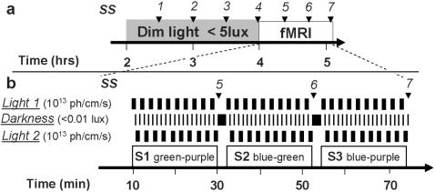
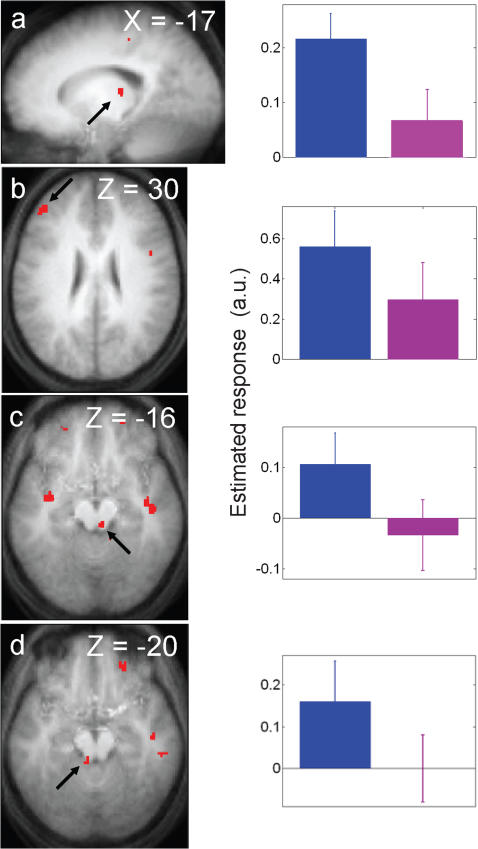
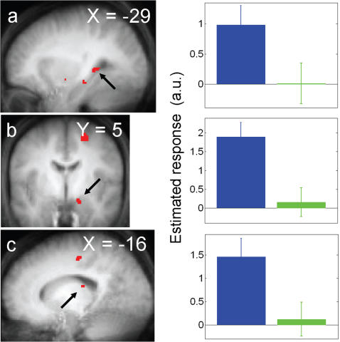
Methodology/principal findings: We exposed 15 participants to short duration (50 s) monochromatic violet (430 nm), blue (473 nm), and green (527 nm) light exposures of equal photon flux (10(13)ph/cm(2)/s) while they were performing a working memory task in fMRI. At light onset, blue light, as compared to green light, increased activity in the left hippocampus, left thalamus, and right amygdala. During the task, blue light, as compared to violet light, increased activity in the left middle frontal gyrus, left thalamus and a bilateral area of the brainstem consistent with activation of the locus coeruleus.
Conclusion/significance: These results support a prominent contribution of melanopsin-expressing retinal ganglion cells to brain responses to light within the very first seconds of an exposure. The results also demonstrate the implication of the brainstem in mediating these responses in humans and speak for a broad involvement of light in the regulation of brain function.
Conflict of interest statement
Figures
References
-
- Dijk DJ, Lockley SW. Integration of human sleep-wake regulation and circadian rhythmicity. J Appl Physiol. 2002;92:852–862. - PubMed
-
- Cajochen C, Munch M, Kobialka S, Krauchi K, Steiner R, et al. High sensitivity of human melatonin, alertness, thermoregulation, and heart rate to short wavelength light. J Clin Endocrinol Metab. 2005;90:1311–1316. - PubMed
-
- Lockley SW, Brainard GC, Czeisler CA. High sensitivity of the human circadian melatonin rhythm to resetting by short wavelength light. J Clin Endocrinol Metab. 2003;88:4502–4505. - PubMed
-
- Lockley SW, Evans EE, Scheer FAJL, Brainard GC, Czeisler CA, et al. Short-wavelength sensitivity for the direct effects of light on alertness, vigilance, and the waking electroencephalogram in humans. Sleep. 2006;29:161–168. - PubMed
-
- Munch M, Kobialka S, Steiner R, Oelhafen P, Wirz-Justice A, et al. Wavelength-dependent effects of evening light exposure on sleep architecture and sleep EEG power density in men. Am J Physiol Regul Integr Comp Physiol. 2006;290:R1421–1428. - PubMed
