Review
. 2007 Dec;85(12):1331-8.
doi: 10.1007/s00109-007-0283-1. Epub 2007 Nov 28.

Harnessing hypoxic adaptation to prevent, treat, and repair stroke

Affiliations
Review

Harnessing hypoxic adaptation to prevent, treat, and repair stroke

Rajiv R Ratan et al. J Mol Med (Berl). 2007 Dec.

Abstract

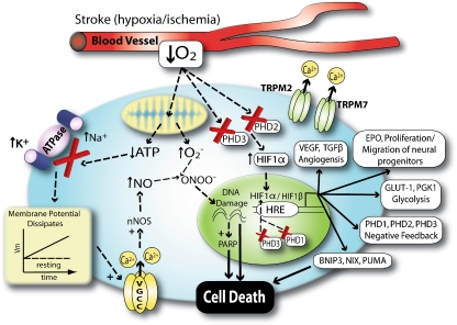
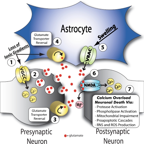
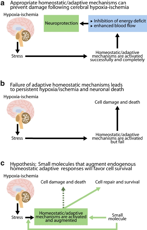
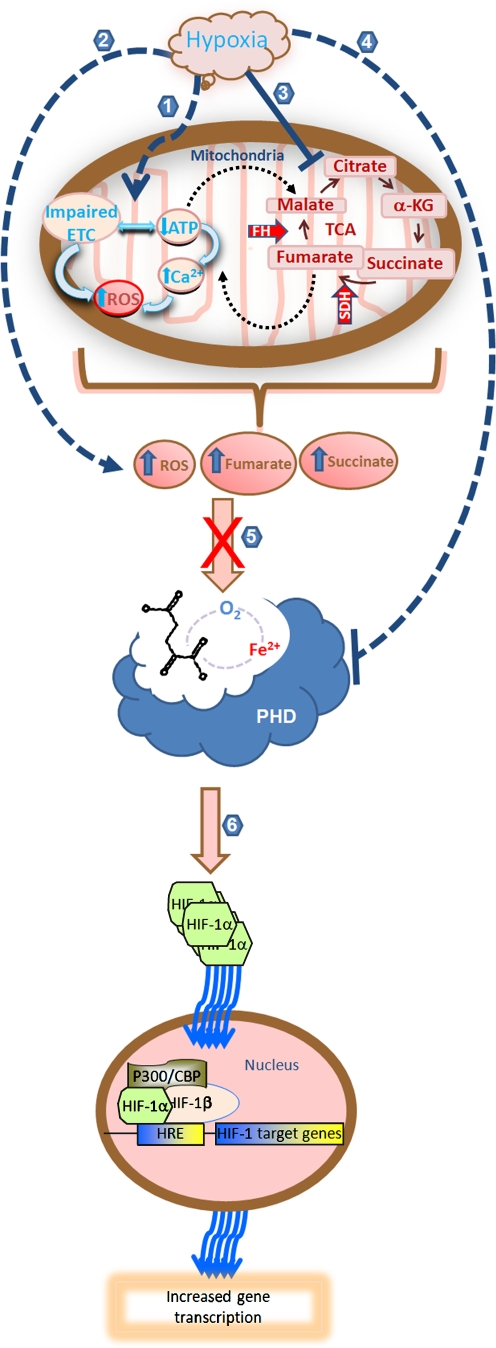
The brain demands oxygen and glucose to fulfill its roles as the master regulator of body functions as diverse as bladder control and creative thinking. Chemical and electrical transmission in the nervous system is rapidly disrupted in stroke as a result of hypoxia and hypoglycemia. Despite being highly evolved in its architecture, the human brain appears to utilize phylogenetically conserved homeostatic strategies to combat hypoxia and ischemia. Specifically, several converging lines of inquiry have demonstrated that the transcription factor hypoxia-inducible factor-1 (HIF1-1) mediates the activation of a large cassette of genes involved in adaptation to hypoxia in surviving neurons after stroke. Accordingly, pharmacological or molecular approaches that engage hypoxic adaptation at the point of one of its sensors (e.g., inhibition of HIF prolyl 4 hydroxylases) leads to profound sparing of brain tissue and enhanced recovery of function. In this review, we discuss the potential mechanisms that could subserve protective and restorative effects of augmenting hypoxic adaptation in the brain. The strategy appears to involve HIF-dependent and HIF-independent pathways and more than 70 genes and proteins activated transcriptionally and post-transcriptionally that can act at cellular, local, and system levels to compensate for oxygen insufficiency. The breadth and depth of this homeostatic program offers a hopeful alternative to the current pessimism towards stroke therapeutics.


Figures

Fig. 1
Fig. 1
Neuronal hypoxia leads to calcium overload and production of free radicals. Stroke is associated with a decrease in cerebral blood flow to the brain. The consequent loss of metabolic fuels leads to failure of sodium pumps leading to an intracellular accumulation of sodium and calcium, depolarization, and activation of voltage sensitive and ligand gated [N-methyl-d-aspartate (NMDA)] ion channels. Increased in calcium in microdomains near the NMDA receptor leads to activation of neuronal nitric oxide synthase. Global dysregulation of calcium in the neuron leads to mitochondrial overload and superoxide production. Nitric oxide and superoxide combine to form peroxynitrite. Peroxynitrite can damage DNA leading to PARP activation and consumption of NAD+. It can also activate TRPM2/7 channels leading to further calcium dysregulation. Hypoxia is sensed by decreased activity of HIF prolyl 4 hydroxylases that can lead to activation genetic responses capable of compensating for the sentinel metabolic stress (decreased cerebral blood flow). Decrease HIF PHD activity can also prevent death via HIF-independent pathways. Acidosis and ROS can also combine with HIF regulated prodeath proteins to trigger cell death
Fig. 2
Fig. 2
Contribution of neurons and astrocytes in mediating excitotoxic neuronal death. 1 Loss of ATP in ischemia leads to inhibition of the Na+/K+ATPase and subsequent collapse of normal ionic gradients. 2 In turn, neuronal membrane depolarization activates voltage sensitive Ca2+ channels, which increase intracellular Ca2+ and stimulate vesicular glutamate release. Severe loss of ionic gradients found in certain ischemic regions may also lead to the reversal of 3 neuronal specific and 4 astrocyte specific glutamate transporters, which in the reverse mode act to release glutamate into the extracellular space. 5 Cell swelling in cerebral ischemia, which is mainly localized to astrocytes, likely activates swelling sensitive anion channels, referred to as volume regulated anion channels (VRACs). VRACs, which are permeable to organic osmolytes, contribute to glutamate release predominantly in the ischemic penumbra. 6 Glutamate regulated NMDA receptors (NMDA-R) are activated by (1) extracellular glutamate and (2) release of Mg2+ from its pore after membrane depolarization (in part due to activation of glutamate regulated AMPA receptors, not shown). 7 NMDA-Rs are permeable to Ca2+ and as such, overabundant NMDA-R activation leads to an intracellular Ca2+ overload. This increase in intracellular Ca2+ then contributes to neuronal death via several mechanisms
Fig. 3
Fig. 3
Drugs that augment endogenous homeostatic mechanisms will more effectively neutralize the heterogeneity inherent in stroke pathophysiology. As these pathways are already used by the body, their activation can occur with decreased threat of toxicity. The term “homeostasis” was coined by Walter Canon in the early twentieth century. It refers to the innate tendency of organisms to mobilize adaptive responses physiological and pathological perturbations that ultimately return the system to a set point that is consistent with survival. a The experimental paradigm of “ischemic preconditioning” has shown that a short, sublethal exposure to hypoxia, or hypoxia-ischemia induces homeostatic responses that make the organism “immune” or “tolerant” to a lethal ischemic insult. Mechanistic studies have revealed that tolerance is the consequence of activation of pre-existing proteins and de novo gene expression. b According to this model, stroke can be conceptualized as a failure of homeostasis. Consequently, neurons die and the brain is permanently damaged. c By extension, small molecules that engage homeostatic mechanisms designed to alleviate hypoxia/ischemia early or enhance their activation should tip the balance away from cell death and toward survival and repair. Such small molecules are currently being developed and represent a new generation of stroke therapies
Fig. 4
Fig. 4
HIF prolyl 4 hydroxylases sense hypoxia and transduce a critical insufficiency in oxygen in the brain into transcriptional and post-transcriptional signal changes that mediate protection and repair. Hypoxia regulates the activity of HIF PHDs via direct or indirect mechanisms; production of peroxide via reduction in mitochondrial ATP production and electron transport chain (ETC) impairment (1, 2); accumulation of the tricarboxylic cycle (TCA) intermediates succinate and fumarate (3); or direct inhibition of the activity of PHDs due to lack of oxygen (4). Accumulation of hydrogen peroxide, succinate or fumarate can inhibits the activity of PHDs by competing with 2-oxoglutarate or by oxidizing the active site iron (5). Among its numerous downstream effects, inhibition of HIF PHD activity leads to stabilization of HIF-1α. Stabilized HIF-1α dimerizes with HIF-1β in the nucleus and increases gene transcription (6)
Fig. 5
Fig. 5
Adaptation to hypoxia-cell fate and beyond. Expression of HIF in neurons leads to the constitutive expression of proteins associated with cell death (BNIP3, NIX, and PUMA) and cell survival (VEGF, glycolytic enzymes, Epo, and p21 waf1/cip1). Similar prodeath gene expression is found in neurons exposed to hypoxia or hypoxia mimetics despite the absence of cell death. It appears the oxygen “starved” neurons have stepped to the edge of the cliff. If during the ensuing hours to days the neuron becomes acidotic or oxidized, then prodeath proteins such as BNIP3 undergo a conformational change, insertion into the mitochondrial membrane, release of apoptotic effectors, and death. By contrast, if the survival genes are effective in neutralizing the hypoxic stress (e.g., no acidosis or oxidative stress), then the death genes never get activated. Our studies indicate that antioxidants, short interfering RNAs to BNIP3 or inhibitors of the HIF prolyl 4 hydroxylases tip the balance toward survival (away from the cliff)

References

    1. {'text': '', 'ref_index': 1, 'ids': [{'type': 'DOI', 'value': '10.1002/ana.21127', 'is_inner': False, 'url': 'https://doi.org/10.1002/ana.21127'}, {'type': 'PubMed', 'value': '17420989', 'is_inner': True, 'url': 'https://pubmed.ncbi.nlm.nih.gov/17420989/'}]}
    2. Savitz SI, Fisher M (2007) Future of neuroprotection for acute stroke: in the aftermath of the SAINT trials. Ann Neurol 61:396–402 - PubMed
    1. {'text': '', 'ref_index': 1, 'ids': [{'type': 'PubMed', 'value': '4723697', 'is_inner': True, 'url': 'https://pubmed.ncbi.nlm.nih.gov/4723697/'}]}
    2. Sharbrough FW, Messick JM Jr., Sundt TM Jr. (1973) Correlation of continuous electroencephalograms with cerebral blood flow measurements during carotid endarterectomy. Stroke 4:674–683 - PubMed
    1. {'text': '', 'ref_index': 1, 'ids': [{'type': 'PubMed', 'value': '2448321', 'is_inner': True, 'url': 'https://pubmed.ncbi.nlm.nih.gov/2448321/'}]}
    2. Naritomi H, Sasaki M, Kanashiro M, Kitani M, Sawada T (1988) Flow thresholds for cerebral energy disturbance and Na+ pump failure as studied by in vivo 31P and 23Na nuclear magnetic resonance spectroscopy. J Cereb Blood Flow Metab 8:16–23 - PubMed
    1. {'text': '', 'ref_index': 1, 'ids': [{'type': 'DOI', 'value': '10.1016/0002-9343(81)90834-2', 'is_inner': False, 'url': 'https://doi.org/10.1016/0002-9343(81)90834-2'}, {'type': 'PubMed', 'value': '6112879', 'is_inner': True, 'url': 'https://pubmed.ncbi.nlm.nih.gov/6112879/'}]}
    2. Gibson GE, Pulsinelli W, Blass JP, Duffy TE (1981) Brain dysfunction in mild to moderate hypoxia. Am J Med 70:1247–1254 - PubMed
    1. {'text': '', 'ref_index': 1, 'ids': [{'type': 'PubMed', 'value': '3247067', 'is_inner': True, 'url': 'https://pubmed.ncbi.nlm.nih.gov/3247067/'}]}
    2. Hossmann KA, Ophoff BG, Csiba L, Paschen W (1988) Regional pH and electrolyte homeostasis of cat brain after prolonged ischemia. Neurochem Pathol 9:127–137 - PubMed

Publication types

MeSH terms