Comparative Study
. 2008 May;215(2):562-73.
doi: 10.1002/jcp.21338.

ESE-1 is a potent repressor of type II collagen gene (COL2A1) transcription in human chondrocytes

Affiliations
Comparative Study

ESE-1 is a potent repressor of type II collagen gene (COL2A1) transcription in human chondrocytes

Haibing Peng et al. J Cell Physiol. 2008 May.

Abstract

The epithelium-specific ETS (ESE)-1 transcription factor is induced in chondrocytes by interleukin-1beta (IL-1beta). We reported previously that early activation of EGR-1 by IL-1beta results in suppression of the proximal COL2A1 promoter activity by displacement of Sp1 from GC boxes. Here we report that ESE-1 is a potent transcriptional suppressor of COL2A1 promoter activity in chondrocytes and accounts for the sustained, NF-kappaB-dependent inhibition by IL-1beta. Of the ETS factors tested, this response was specific to ESE-1, since ESE-3, which was also induced by IL-1beta, suppressed COL2A1 promoter activity only weakly. In contrast, overexpression of ETS-1 increased COL2A1 promoter activity and blocked the inhibition by IL-1beta. These responses to ESE-1 and ETS-1 were confirmed using siRNA-ESE1 and siRNA-ETS1. In transient cotransfections, the inhibitory responses to ESE-1 and IL-1beta colocalized in the -577/-132 bp promoter region, ESE-1 bound specifically to tandem ETS sites at -403/-381 bp, and IL-1-induced binding of ESE-1 to the COL2A1 promoter was confirmed in vivo by ChIP. Our results indicate that ESE-1 serves a potent repressor function by interacting with at least two sites in the COL2A1 promoter. However, the endogenous response may depend upon the balance of other ETS factors such as ETS-1, and other IL-1-induced factors, including EGR-1 at any given time. Intracellular ESE-1 staining in chondrocytes in cartilage from patients with osteoarthritis (OA), but not in normal cartilage, further suggests a fundamental role for ESE-1 in cartilage degeneration and suppression of repair.


Figures

Fig. 1
Fig. 1
Potential ESE-1 binding sites in the COL2A1 promoter. The structure of the COL2A1 regulatory region spanning –577 to +3,426 bp is shown at top with potential ETS binding site indicated with arrows. The previously identified Sp1, Egr-1/Sp1, E-box, and TATA-box sites are represented schematically within the –577/+125 bp promoter. The oligonucleotides that were used for EMSAs are listed below with putative ETS binding sites underlined (left) and the corresponding mutant oligonucleotides (right).
Fig. 2
Fig. 2
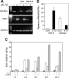
The suppression of COL2A1 gene expression by IL-1β requires NF-κB and is associated with NF-κB-dependent induction of ESE-1 mRNA. A: Cultures of the immortalized human chondrocyte cell line, C-28/I2,were infected with adenovirus expressing IκB (adv-IkB) or, as a null control, adv-βgal, incubated for 4 h in serum-containing medium, changed to serum-free medium containing 1% Nutridoma, and treated with IL-1β (500 pg/ml) for 18 h. Total RNA was extracted and analyzed by RT-PCR using specific primers to detect COL2A1, ESE1, and GAPDH mRNAs. B: Primary cultures of human adult articular chondrocytes at day 5 after isolation from the cartilage were treated with IL-1β for 18h, and ESE1 and COL2A1 mRNA were analyzed by real-time RT-PCR. C: The C-28/I2 cells were cultured in serum-containing culture medium for 5 days, and confluent cultures were changed to serum-free medium containing 1% Nutridoma 24 h before treatment with IL-1β for the times indicated. Emetine (10 μg/ml) was also added to the indicated cultures. Total RNA extracts were analyzed for ESE1 mRNA expression by real-time PCR.
Fig. 3
Fig. 3
ESE-1 decreases COL2A1 promoter activity and enhances IL-1β-dependent inhibition. The C-28/I2 chondrocytes were cotransfected with pGL2B or with pGL2-COL2 together with pCI empty vector or with (A) increasing amounts of pCI-ESE1 (50, 100, and 200 ng/ml), (B) pCI-ESE1 (50 ng/ml) in the absence or presence of increasing amounts of dominant-negative (DN)-ESE1. C: The C-28/I2 cells were cotransfected with pGL2-COL2 and pcDNA3.1 (empty vector) or pcDNA-ESE1 together with siRNA-Ctrl (control) or siRNA-ESE1. Incubations were for 24 h followed by treatment with IL-1β (500 pg/ml) for a further 18 h.
Fig. 4
Fig. 4
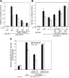
Comparison of ESE-1 with other ETS family members. C-28/I2 cells were cotransfected with pGL2-COL2 and pCI or expression plasmids encoding (A) various ETS family members (100 ng/well in 12-well plates) or (B) ESE-1 (100 ng/ml) or ETS-1 (50 and 100 ng/ml). C: The C-28/I2 cells were cotransfected with pGL2-COL2 and pcDNA3.1 (empty vector) or pcDNA-ETS1 together with siRNA-Ctrl (control) or siRNA-ETS1. The cells were incubated for 24 h prior to the addition of IL-1β for a further 18 h, cell harvest, and luciferase assay.
Fig. 5
Fig. 5
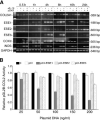
IL-1β induces ESE-1 and ESE-3 coordinately with the suppression of COL2A1 mRNA but ESE-1 inhibits COL2A1 promoter activity more effectively than ESE-3. A: The C-28/I2 cells were cultured as describe in Figure 2C and treated with IL-1β and cycloheximide alone or together for the times indicated. Total RNA extracts were analyzed by semi-quantitative RT-PCR for COL2A1, ESE1, ESE2, ESE3, and GAPDH mRNAs. COX2 and NOS2 mRNAs were also assayed as positive controls. B: The C-28/I2 cells were cotransfected with pGL2-COL2 together with pCI empty vector or increasing amounts (25, 50, 100, 150, and 200 ng/ml) of pCI-ESE1 or pCI-ESE3.
Fig. 6
Fig. 6
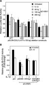
ESE-1 decreases activity of the COL2A1 promoter containing the –577/–132 bp region. A: The C-28/I2 cells were cotransfected with pGL2 constructs containing COL2A1 promoter sequences, –577/+125 bp, –403/+125 bp, –131/+125 bp, and –90/+125 bp, together 200 ng of pCI (filled bar) or 50, 100, or 200 ng/ml of pCI-ESE1. B: The cells were cotransfected with pGL2-COL2 and pcDNA3.1 (empty vector) or expression plasmids encoding ESE-1 (100 ng/ml), DN-ESE1 (50 ng/ml), or DN-ESE1 and DN-Egr1, each at 50 ng/ml. The cultures were then incubated for 24 h to permit expression of recombinant proteins. Some cultures were then treated with IL-1β (empty bar) and the incubations continued for a further 18h. All cultures were harvested at the same time for luciferase assay of reporter gene expression. The results show the means ± SD derived from three separate transfection experiments, each done in triplicate.
Fig. 7
Fig. 7
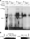
ESE-1 binds to ETS sites E2 and E3 within –403/–381 bp. A: End-labeled double-stranded oligonucleotides containing the ESE-1 consensus (NOS2 promoter) or the E2, E3, and E4 oligonucleotides or mutants (see Fig. 1) were incubated with in vitro translated protein made with empty vector (v) or pCI-ESE1 (E). Competitor oligonucleotides (at 50× and 100× in adjacent lanes) and antibody were added as indicated and as described in the Methods. Similar background binding of in vitro-translated vector or ESE-1 has been reported on other promoter sequences (Brown et al., 2004; Wang et al., 2004; Grall et al., 2005). Note that the third lane of each set shows binding to mutant (mt) E2, E3, and E4 probes. B: Chromatin immunoprecipitations (ChIP) were performed using C-28/I2 cells transfected with pcDNA3-FLAG empty vector (EV) or pcDNA3-FLAG-ESE1 or -DN-ESE1 (left), or they were treated with IL-1β for 6 h (right). Chromatin proteins were crosslinked to DNA with formaldehyde and purified nucleoprotein complexes were immunoprecipitated using anti (α)-FLAG or α-ESE1 antibody. The precipitated DNA fractions were analyzed by PCR for the presence of the COL2A1 promoter region encompassing the ESE-1 binding sites. The input DNA was used as a positive control for the PCR and water was used as negative control (not shown).
Fig. 8
Fig. 8
Immunolocalization of ESE-1 in chondrocytes in vivo associated with osteoarthritis but not in normal articular cartilage and induction of ESE-1 mRNA by IL-1β in primary human articular chondrocytes. Samples of articular cartilage from individuals without apparent joint destruction (normal; a and c) and from patients with osteoarthritis (OA; b and d) were subjected to immunohistochemical staining using an antibody against human ESE-1 or isotype-matched IgG as negative control. Positive staining for ESE-1 appears as a brown color in the cytoplasm and nuclei of chondrocytes in this representative sample of OA cartilage. (Original magnifications, 40×). [Color figure can be viewed in the online issue, which is available at www.interscience.wiley.com.]
Fig. 9
Fig. 9
Mechanism of suppression of COL2A1 promoter activity by IL-1β-induced transcription factors.

Similar articles

Cited by

References

    1. Andriamanalijaona R, Felisaz N, Kim SJ, King-Jones K, Lehmann M, Pujol JP, Boumediene K. Mediation of interleukin-1b-induced transforming growth factor b1 expression by activator protein 4 transcription factor in primary cultures of bovine articular chondrocytes: Possible cooperation with activator protein 1. Arthritis Rheum. 2003;48:1569–1581. - PubMed
    1. Bau B, Gebhard PM, Haag J, Knorr T, Bartnik E, Aigner T. Relative messenger RNA expression profiling of collagenases and aggrecanases in human articular chondrocytes in vivo and in vitro. Arthritis Rheum. 2002;46:2648–2657. - PubMed
    1. Brown C, Gaspar J, Pettit A, Lee R, Gu X, Wang H, Manning C, Voland C, Goldring SR, Goldring MB, Libermann TA, Gravalllese EM, Oettgen P. ESE-1 is a novel transcriptional mediator of angiopoietin-1 expression in the setting of inflammation. J Biol Chem. 2004;279:12794–12803. - PubMed
    1. Chadjichristos C, Ghayor C, Kypriotou M, Martin G, Renard E, Ala-Kokko L, Suske G, de Crombrugghe B, Pujol JP, Galera P. Sp1 and Sp3 transcription factors mediate interleukin-1 b down-regulation of human type II collagen gene expression in articular chondrocytes. J Biol Chem. 2003;278:39762–39772. - PubMed
    1. Czuwara-Ladykowska J, Shirasaki F, Jackers P, Watson DK, Trojanowska M. Fli-1 inhibits collagen type I production in dermal fibroblasts via an Sp1-dependent pathway. J Biol Chem. 2001;276:20839–20848. - PubMed

Publication types

MeSH terms