. 2007 Nov 27;4(11):e328.
doi: 10.1371/journal.pmed.0040328.

An LRP5 receptor with internal deletion in hyperparathyroid tumors with implications for deregulated WNT/beta-catenin signaling

Affiliations

An LRP5 receptor with internal deletion in hyperparathyroid tumors with implications for deregulated WNT/beta-catenin signaling

Peyman Björklund et al. PLoS Med. .

Abstract

Background: Hyperparathyroidism (HPT) is a common endocrine disorder with incompletely understood etiology, characterized by enlarged hyperactive parathyroid glands and increased serum concentrations of parathyroid hormone and ionized calcium. We have recently reported activation of the Wnt signaling pathway by accumulation of beta-catenin in all analyzed parathyroid tumors from patients with primary HPT (pHPT) and in hyperplastic parathyroid glands from patients with uremia secondary to HPT (sHPT). Mechanisms that may account for this activation have not been identified, except for a few cases of beta-catenin (CTNNB1) stabilizing mutation in pHPT tumors.

Methods and findings: Reverse transcription PCR and Western blot analysis showed expression of an aberrantly spliced internally truncated WNT coreceptor low-density lipoprotein receptor-related protein 5 (LRP5) in 32 out of 37 pHPT tumors (86%) and 20 out of 20 sHPT tumors (100%). Stabilizing mutation of CTNNB1 and expression of the internally truncated LRP5 receptor was mutually exclusive. Expression of the truncated LRP5 receptor was required to maintain the nonphosphorylated active beta-catenin level, transcription activity of beta-catenin, MYC expression, parathyroid cell growth in vitro, and parathyroid tumor growth in a xenograft severe combined immunodeficiency (SCID) mouse model. WNT3 ligand and the internally truncated LRP5 receptor strongly activated transcription, and the internally truncated LRP5 receptor was insensitive to inhibition by DKK1.

Conclusions: The internally truncated LRP5 receptor is strongly implicated in deregulated activation of the WNT/beta-catenin signaling pathway in hyperparathyroid tumors, and presents a potential target for therapeutic intervention.


Conflict of interest statement

Competing Interests: The authors declare the following competing interest: A patent is filed (USA) for “LRP5 and LRP6 Receptors in Cancer Treatment,” number WO2005048913.

Figures

Figure 1
Figure 1. An Internally Truncated LRP5 Receptor Is Expressed in Parathyroid Tumors
(A) RT-PCR analysis of RNA from pHPT tumors (n = 20) and sHPT tumors (n = 20) using primers located in exons 9 and 13 of LRP5. The wild-type or truncated LRP5 mRNAs were detected by primary PCR for most of the tumors or with nested PCR using an additional overlapping forward primer. The truncation comprised the last 93 bp of exon 9, all 227 bp of exon 10, and the first 106 bp of exon 11. (B) Nested RT-PCR of normal parathyroid tissue (n = 6), and parathyroid tumor cell line sHPT-1 [8] as marker (top panel). Nested RT-PCR analysis of 17 normal tissues, and the parathyroid tumor pHPT21 as marker (lower panel). (C) Immunoprecipitation followed by Western blot analysis of LRP5. Transiently expressed (HEK293T cells) LRP5 and LRP5Δ666–809 shown as size markers. sHPT-1, human parathyroid tumor cell line [8]; pHPT, pHPT tumor. The lower panel shows additional pHPT tumor samples analyzed on a 5% SDS–polyacrylamide gel, where the proteins separate more clearly. (D) A schematic structure of LRP5 is shown. The truncated mRNA contains an in-frame deletion of LRP5 between amino acids 666 and 809 (Δ666–809), encompassing the third YWTD β-propeller domain. The truncation (Δ666–809) is flanked by imperfect direct repeat sequences (horizontal arrows) with putative cryptic donor (Ac-GTG) and acceptor (AC-cT) RNA splice sites in exons 9 and 11, respectively (arrows). The Δ666–809 is between nucleotide positions 2039 and 2466 of the LRP5 mRNA (GenBank accession no. AF064548).
Figure 2
Figure 2. Specificity and Efficiency of siRNAs Transiently Transfected to the sHPT-1 Parathyroid Tumor Cell Line
(A) Locations of siRNAs and probes for quantitative real-time PCR. tot mRNA PCR probe determines both LRP5wt and LRP5Δ666–809 transcripts. (B) Expression of LRP5wt and LRP5tot mRNA assessed by quantitative real-time PCR. (C) Immunoprecipitation and Western blot analysis of LRP5.
Figure 3
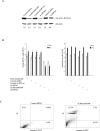
Figure 3. Maintained Expression of the Internally Truncated LRP5 Receptor Is Required for Accumulation of Nonphosphorylated β-Catenin and Continued Cell Growth
(A) Western blot analysis of active β-catenin [25], 60 h after transfection. The β-catenin–actin signal ratio is shown. (B) Effects on sHPT-1 cell growth. HeLa cells were used as control for toxic effects. *p < 0.05. (C) Flow cytometry analysis of sHPT-1 cells at 84 h after transfection after staining with annexin V–FITC and propidium iodide. Accumulation of dead cells in the upper left quadrant; population of late apoptotic cells (upper right quadrant) and early apoptotic cells (lower right quadrant).
Figure 4
Figure 4. The Internally Truncated LRP5 Receptor Regulates β-Catenin–Driven Transcription
(A) Transient cotransfections of FOPFLASH or TOPFLASH TCF/β-catenin reporters, the CMV-LacZ reference plasmid, and indicated siRNAs to sHPT-1 cells (left panel). FOPFLASH contains mutated binding sites for TCF factors, while TOPFLASH does not [22]. Luciferase activities were normalized to β-galactosidase activities. Effects on endogenous MYC expression in sHPT-1 cells (right panel). *p < 0.05. (B) Western blot analysis of V5-tagged LRP5 and LRP5Δ666–809 transiently transfected to HEK293T cells. LRP5 and LRP5Δ666–809 were expressed at similar levels. (C) Western blot analysis of cytosolic nonphosphorylated active β-catenin in HEK293T cells, 24 h after transfection. (D) Endogenous MYC expression in transfected HEK293T cells. *p < 0.05. (E) ChIP of the MYC promoter in transfected HEK293T cells. An anti-active–β-catenin monoclonal antibody was used [25].
Figure 5
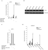
Figure 5. WNT3 Ligand and the Internally Truncated LRP5 Receptor Strongly Activates Transcription of the TOPFLASH TCF/β-Catenin Luciferase Reporter with Impaired DKK1 Inhibition
(A) sHPT-1 cells cotransfected with TOPFLASH, LRP5wt, or LRP5Δ666–809 expression vectors and CMV-LacZ reference plasmid, followed by incubation in WNT1, WNT3, or WNT3A conditioned medium (CM). CM was from HEK293T cells transfected transiently with expression vectors for the various WNT ligands. The 11-fold (Figure 4A) endogenous β-catenin activity is set to 1 (unstimulated, empty vector). (B) Representative RT-PCR analysis of RNA from three pHPT and three sHPT tumors using primers for WNT3. No expression of WNT1 was detected by the conditions used [21]. (C) Cotransfection of TOPFLASH and CMV-LacZ reference plasmid to sHPT-1 cells. Incubation in WNT3 and DKK1 CM. (D) Cotransfection of TOPFLASH, CMV-LacZ reference plasmid, and LRP5wt or LRP5Δ666–809 expression vectors to HEK293T cells followed by incubation in WNT1, WNT3, or DKK1 CM. HEK293T cells do not express the internally truncated LRP5 receptor.
Figure 6
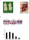
Figure 6. siRNAs to the Internally Truncated LRP5 Receptor Inhibit Tumor Growth in Xenografted SCID Mice
(A) Injection of sHPT-1 cells pretransfected for 24 h with siLRP5Δ666–809 or control siRNA. Arrows indicate representative parathyroid tumor growth at site of transplantation. (B) Parathyroid tumors from SCID mice (n = 35) injected with sHPT-1 cells pretransfected for 24 h with the indicated siRNAs were excised and weighed. *p < 0.05. The animals were monitored every day and killed after 8–9 wk.

References

    1. Marx SJ. Hyperparathyroid and hypoparathyroid disorders. N Engl J Med. 2000;343:1863–1875. - PubMed
    1. Arnold A, Shattuck TM, Mallya SM, Krebs LJ, Costa J, et al. Molecular pathogenesis of primary hyperparathyroidism. J Bone Miner Res. 2002;17(Suppl 2):N30–N36. - PubMed
    1. Åkerström G, Hellman P. Primary hyperparathyroidism. Curr Opin Oncol. 2004;16:1–7. - PubMed
    1. Åkerström G, Hellman P, Hessman O, Segersten U, Westin G. Parathyroid glands in calcium regulation and human disease. Ann N Y Acad Sci. 2005;1040:53–58. - PubMed
    1. Björklund P, Åkerström G, Westin G. Accumulation of nonphosphorylated beta-catenin and c-myc in primary and uremic secondary hyperparathyroid tumors. J Clin Endocrinol Metab. 2007;92:338–344. - PubMed

Publication types