. 2008 Mar 15;410(3):585-94.
doi: 10.1042/BJ20071505.

EGF and amphiregulin differentially regulate Cbl recruitment to endosomes and EGF receptor fate

Affiliations

EGF and amphiregulin differentially regulate Cbl recruitment to endosomes and EGF receptor fate

Kathryn A Stern et al. Biochem J. .

Abstract

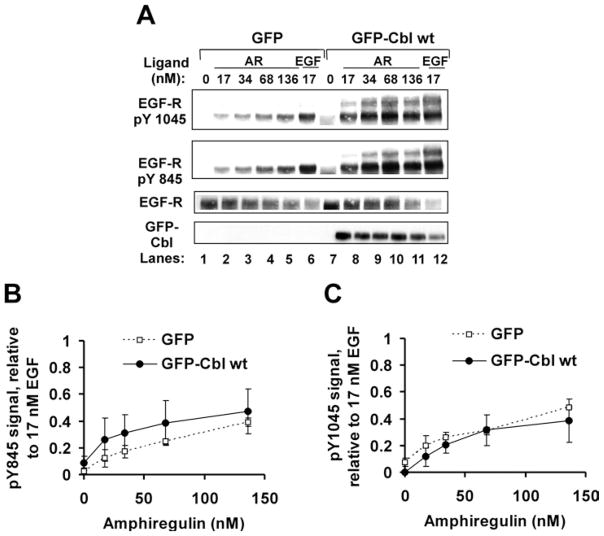
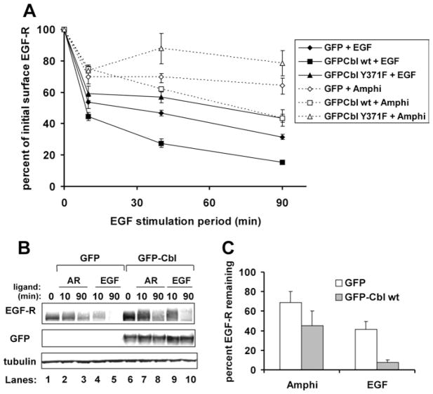
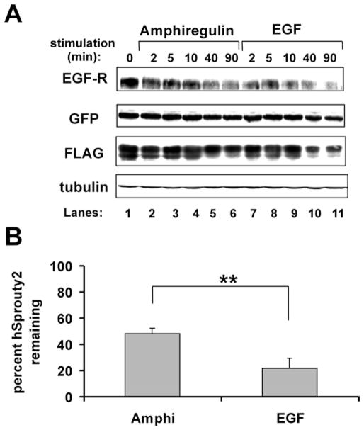
EGF-R [EGF (epidermal growth factor) receptor] ligands can promote or inhibit cell growth. The biological outcome of receptor activation is dictated, at least in part, by ligand-specified patterns of endocytic trafficking. EGF-R trafficking downstream of the ligands EGF and TGF-alpha (transforming growth factor-alpha) has been investigated extensively. However, less is known about EGF-R fates induced by the ligands BTC (betacellulin) and AR (amphiregulin). We undertook comparative analyses to identify ligand-specific molecular events that regulate EGF-R trafficking and degradation. EGF (17 nM) and BTC (8.5 nM) induced significant EGF-R degradation, with or without ectopic expression of the ubiquitin ligase Cbl. Human recombinant AR (17 nM) failed to affect receptor degradation in either case. Notably, levels of ligand-induced EGF-R ubiquitination did not correlate strictly with receptor degradation. Dose-response experiments revealed that AR at a saturating concentration was a partial agonist at the EGF-R, with approx. 40% efficacy (relative to EGF) at inducing receptor tyrosine phosphorylation, ubiquitination and association with Cbl. EGF-R down-regulation and degradation also were compromised upon cell stimulation with AR (136 nM). These outcomes correlated with decreased degradation of the Cbl substrate and internalization inhibitor hSprouty2. Downstream of the hSprouty2 checkpoint in AR-stimulated cells, Cbl-free EGF-R was incorporated into endosomes from which Cbl-EGF-R complexes were excluded. Our results suggest that the AR-specific EGF-R fate results from decreased hSprouty2 degradation and reduced Cbl recruitment to underphosphorylated EGF-R, two effects that impair EGF-R trafficking to lysosomes.


Figures

Figure 1
Figure 1. Equimolar concentrations of AR and EGF induce different levels of EGF-R ubiquitination, phosphorylation and degradation
HEK-293 cells were transiently transfected with cDNA encoding EGF-R (0.05 μg) and either GFP (3 μg) or GFP–Cbl WT (4 μg). At 48 h post-transfection, cells were serum-starved and incubated without (0 nM) or with AR (17 nM), BTC (8.5 nM) or EGF (17 nM) for 10, 40 or 90 min. Cell lysate proteins and immunoprecipitates (IP) were gel-resolved, transferred on to PVDF and immunoblotted (IB) with the antibodies indicated. (A) Immunoblotting results from a representative experiment. Top arrow: ubiquitinated EGF-R; middle arrow, non-ubiquitinated EGF-R; bottom arrow, GFP–Cbl; *, phosphorylated Hrs, a marker for EGF-R complex trafficking to early endosomes. (B) Quantitative analysis of three independent experiments, showing the amount of total cellular EGF-R remaining after receptor stimulation by the ligands (Amphi, amphiregulin). Results are means ± S.D. A two-tailed Student’s t test with α=0.05 determined that GFP–Cbl expression significantly affected EGF-R degradation only in the case of receptors activated by EGF (**).
Figure 2
Figure 2. Human recombinant AR is a partial agonist at the EGF-R with approx. 40 % efficacy for total EGF-R tyrosine phosphorylation, ubiquitination and co-precipitation of Cbl
HEK-293 cells were transiently transfected with cDNA encoding EGF-R (0.05 μg) and either GFP (3 μg) or GFP–Cbl WT (4 μg). The cells were serum-starved and then harvested without further treatment (0 nM ligand) or following treatment for 10 min with the indicated ligands at the concentration shown. (A) Immunoblotting results from a representative experiment. Anti-EGFR immunoprecipitates and cell lysate samples were resolved by SDS/PAGE, transferred on to PVDF and immunoblotted with the antibodies shown. Asterisks (*) mark the position of ubiquitinated EGF-R. (BD) Quantitative analysis of three independent experiments. Results are means ± S.D. (B) Total induced EGF-R tyrosine phosphorylation (pY) reaches a plateau upon cell stimulation with approx. 34 nM AR. The phosphotyrosine content of immunoprecipitated receptors was normalized to EGF-R levels. (C) Total induced EGF-R ubiquitination reaches a plateau upon cell stimulation with approx. 68 nM AR. The ubiquitin content of immunoprecipitated receptors was normalized to the total amount of EGF-R present. (D) Cbl co-precipitation (co-IP) with EGF-R reaches a plateau upon cell stimulation with approx. 68 nM AR. The amount of EGF-R-associated Cbl was normalized to the total amount of EGF-R present.
Figure 3
Figure 3. AR is a partial agonist for site-specific EGF-R tyrosine phosphorylation
HEK-293 cells were transiently transfected with cDNA encoding EGF-R (0.05 μg) and GFP–Cbl WT (4 μg) or GFP (3 μg). At 48 h post-transfection, cells were serum-starved and incubated without (0) or with EGF (17 nM) or AR (17–136 nM) for 10 min. Cell lysate proteins (100 μg per lane) were gel-resolved, transferred on to PVDF membrane and immunoblotted with the indicated antibodies. (A) Immunoblotting results from a representative experiment. (B, C) Quantitative analysis of phospho-Tyr845 and phospho-Tyr1045 levels in treated cells. In each case, the phosphotyrosine signal was normalized to the corresponding EGF-R level. Results are means ± S.D. for three independent experiments, except in (C), ● (two independent experiments).
Figure 4
Figure 4. Relative to EGF treatment, AR treatment impairs EGFR down-regulation and degradation
HEK-293 cells were transiently transfected with cDNA encoding EGF-R WT (0.05 μg) and GFP (3.2 μg), GFP–Cbl WT (4 μg) or GFP–Cbl Y371F (4 μg). (A) Down-regulation assay. At 48 h post-transfection, the cells were serum-starved and incubated at 37 °C without (0 min) or with EGF (17 nM) or AR (136 nM) for 10, 40 or 90 min. Cells were harvested intact on ice, plated in triplicate and stained with anti-EGF-R, anti-Syk (isotype-matched negative control) or anti-MHC Class I (isotype-matched positive control) antibodies. This was followed by incubation with phycoerythrin-conjugated secondary antibody and paraformaldehyde fixation. The surface phycoerythrin signals were analysed by flow cytometry for 5000 GFP-positive cells per sample. Surface receptor levels for each sample were determined by subtracting the mean fluorescence index of the anti-Syk-stained cells from the MFI of the matched anti-EGF-R-stained cells. For each transfection condition, the surface EGFR levels are expressed as a percentage of the EGF-R signal of unstimulated, matched transfection plates. Results are means ± S.D. for three independent experiments. (B) EGF-R degradation assay. A 100 μg cell lysate protein sample was loaded per lane, gel-resolved, transferred on to PVDF and immunoblotted as indicated. Results shown are from a representative experiment employing 17 nM EGF and 136 nM AR. (C) Quantitative analysis of three independent degradation experiments, showing the average amount of total cellular EGF-R remaining after 90 min of receptor stimulation by ligand. Results are means ± S.D.
Figure 5
Figure 5. hSpry2 degradation is impaired in AR-treated cells
HEK-293 cells were transiently transfected with cDNA encoding EGF-R (0.05 μg), GFP–Cbl WT (4 μg) and FLAG–hSprouty2 (2 μg). At 48 h post-transfection, cells were serum-starved and incubated without (0 min) or with AR (136 nM) or EGF (17 nM) for 2, 5, 10, 40 or 90 min. (A) Immunoblotting results from a representative experiment. A 100 μg cell lysate protein sample was loaded per lane, gel-resolved, transferred on to PVDF and immunoblotted as indicated. (B) Quantitative analysis of three independent degradation experiments, showing the average amount of total cellular hSprouty2 remaining after 90 min of receptor stimulation by ligand. Results are means ± S.D. A two-tailed Student’s t test with α=0.05 determined that a significantly different amount of hSprouty2 degradation was effected by AR compared with EGF (**).
Figure 6
Figure 6. AR treatment leads to the formation of EGF-R-positive vesicles that lack associated GFP–Cbl
COS-7 cells were transiently transfected with cDNA encoding GFP–Cbl WT (4 μg). At 48 h post-transfection, cells were serum-starved and incubated without ligand (0 min) or with AR (136 nM) or EGF (17 nM) for 5 or 25 min. Following stimulation, the cells were paraformaldehyde-fixed, permeabilized and stained with anti-EGF-R antibody. Scale bar, 20 μm. Images shown are representative results from one experiment. Three independent experiments were performed, with evaluation of more than 20 cells per ligand condition per experiment.

References

    1. Luetteke NC, Qiu TH, Fenton SE, Troyer KL, Riedel RF, Chang A, Lee DC. Targeted inactivation of the EGF and amphiregulin genes reveals distinct roles for EGF receptor ligands in mouse mammary gland development. Development. 1999;126:2739–2750. - PubMed
    1. Xian CJ, Li L, Deng YS, Zhao SP, Zhou XF. Lack of effects of transforming growth factor-α gene knockout on peripheral nerve regeneration may result from compensatory mechanisms. Exp Neurol. 2001;172:182–188. - PubMed
    1. Abbott BD, Lin TM, Rasmussen NT, Albrecht RM, Schmid JE, Peterson RE. Lack of expression of EGF and TGF-α in the fetal mouse alters formation of prostatic epithelial buds and influences the response to TCDD. Toxicol Sci. 2003;76:427–436. - PubMed
    1. Troyer KL, Luetteke NC, Saxon ML, Qiu TH, Xian CJ, Lee DC. Growth retardation, duodenal lesions, and aberrant ileum architecture in triple null mice lacking EGF, amphiregulin, and TGF-α. Gastroenterology. 2001;121:68–78. - PubMed
    1. Luetteke NC, Qiu TH, Peiffer RL, Oliver P, Smithies O, Lee DC. TGFα deficiency results in hair follicle and eye abnormalities in targeted and waved-1 mice. Cell. 1993;73:263–278. - PubMed

Publication types

MeSH terms