. 2008 Jan;134(1):192-203.
doi: 10.1053/j.gastro.2007.10.022. Epub 2007 Oct 17.

Epithelial: lamina propria lymphocyte interactions promote epithelial cell differentiation

Affiliations

Epithelial: lamina propria lymphocyte interactions promote epithelial cell differentiation

Stephanie Dahan et al. Gastroenterology. 2008 Jan.

Abstract

Background & aims: Intestinal lymphoepithelial interactions occur in the epithelium and the subepithelial space. We asked whether normal, Crohn's disease (CD), or ulcerative colitis (UC) lamina propria lymphocytes (LPL) could promote intestinal epithelial cell (IEC) growth and differentiation.

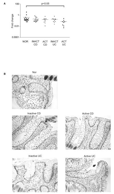
Methods: T84 cells were cocultured with isolated LPL. IECs were then lysed and subjected to measurement of intestinal alkaline phosphatase (IAP) activity; Western blot analysis for MAPK and Akt activation; and real-time polymerase chain reaction to assess caudal-related homeoprotein 2 (CDX2) messenger RNA (mRNA) levels. Tissue sections were immunostained for evidence of mitogen-activated protein kinase (MAPK) and phosphatidylinositol 3-kinase (PI3K) activation, CDX2, and IAP; and CDX2 mRNA expression was assessed in human colonic biopsy specimens.

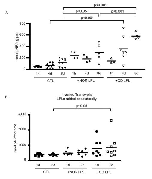
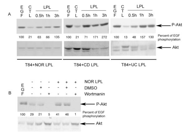
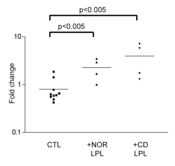
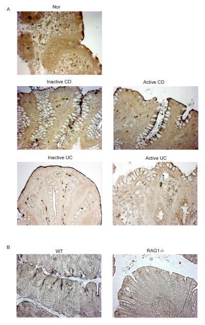
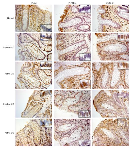
Results: IAP activity was increased in T84 cells cocultured for 8 days with normal LPL (P < .05) and even greater with CD LPL (P < .001). Crypt IECs in active CD mucosa expressed IAP ex vivo. Phospho-MAPK (extracellular signal-regulated kinase 1/2, p38, and c-Jun-N-terminal kinase) and phospho-Akt were seen as early as 30 minutes after coculture. MAPK activation was greatest in T84 cells cocultured with CD LPL. There was a specific increase in Phospho-p38 MAPK and Phospho-Akt staining in the nuclei of crypt IECs in active vs inactive CD, normal mucosa, and UC mucosa. CDX2 mRNA expression was increased in CD LPL cocultured T84 cells, which did not correlate with CDX2 protein localization ex vivo.

Conclusions: There is cross talk between LPL and IECs, which leads to IEC differentiation. The differentiation is accelerated in CD mucosa.


Figures

Figure 1
Figure 1
A. IAP activity in T84 cells co-cultured with Nor or CD LPL for 1 hour, 4 or 8 days. Adherent cells were co-cultured with freshly isolated LPL at a ratio of 1 IEC : 1 LPL. After removing the LPL, the IEC line was lysed and IAP activity (nmol pNP/mg prot) was measured by a colorimetric enzymatic assay and normalized to the protein content in each sample. B. IAP activity in inverted polarized T84 cells co-cultured with Nor or CD LPL for 1 or 2 days. Adherent cells were co-cultured and IAP activity determiend as described in 1A.
Figure 2
Figure 2
PI3K activation in T84 cells co-cultured with LPL. A. Immunoblotting for P-Akt and Akt in lysates obtained from T84 cells co-cultured with freshly isolated Nor, CD or UC LPL for 30 min to 3 hours. The CTL (control) lane contains T84 alone. EGF (10 nM; 15min) was used as a positive control. B. Immunoblotting for P-Akt and Akt in lysates obtained from T84 cells pre-treated for 90 min with DMSO or Wortmannin (100 nM), washed out and co-cultured with freshly isolated Nor LPL for 1 hour. The CTL lanes are as described in 2A. (Representative of 3 experiments). The percentage of Akt activity related to EGF induced activity was quantified by densitometric analysis.
Figure 3
Figure 3
CDX2 mRNA expression in T84 cells co-cultured with Nor or CD LPL for 4 days. Adherent cells were co-cultured as described in 1A. After removing the LPL, T84 mRNA was extracted and quantified by Real Time-PCR. The fold increase in CDX2 mRNA expression was calculated as described in Materials and Methods.
Figure 4
Figure 4
A. IAP staining of paraffin embedded colonic tissue of Nor, inactive and active CD, and inactive and active UC. The slides were examined with a Zeiss Axioskop light microscope at 20X magnification. These data are representative of 4 experiments. B. IAP staining of paraffin embedded colonic tissue from WT and RAG1−/− mice (20X magnification). (Representative of 4 experiments).
Figure 5
Figure 5
Nor, inactive and active CD, and inactive and active UC colonic tissue sections were immunostained using anti-phospho Akt, anti-phospho PTEN, and anti-Cyclin D1 antibodies. The slides were counter-stained with Mayer’s Hematoxylin solution, and examined with a Zeiss Axioskop light microscope at 20X magnification. The insets represent 100X magnification of the crypt epithelial cells to document nuclear localization. (Representative of 4 experiments).
Figure 6
Figure 6
A. CDX2 mRNA expression in surface IECs from Nor, inactive and active CD, and inactive and active UC colonic biopsies. The IECs were isolated, mRNA extracted and quantified by Real Time-PCR. The fold increase of CDX2 mRNA expression was calculated as described in Materials and Methods. B. Colonic tissue sections were immunostained and analyzed as described in Fig 5 using anti-CDX2 antibodies. The insets represent 100X magnification of the surface epithelial cells. (Representative of 4 experiments).

References

    1. Hershberg RM, Mayer LF. Antigen processing and presentation by intestinal epithelial cells - polarity and complexity. Immunol Today. 2000;21:123–8. - PubMed
    1. Dahan S, Roth-Walter F, Arnaboldi P, Agarwal S, Mayer L. Epithelia: lymphocyte interactions in the gut. Immunol Rev. 2007;215:243–53. - PMC - PubMed
    1. Allez M, Brimnes J, Dotan I, Mayer L. Expansion of CD8+ T cells with regulatory function after interaction with intestinal epithelial cells. Gastroenterology. 2002;123:1516–26. - PubMed
    1. Allez M, Brimnes J, Shao L, Dotan I, Nakazawa A, Mayer L. Activation of a unique population of CD8(+) T cells by intestinal epithelial cells. Ann N Y Acad Sci. 2004;1029:22–35. - PubMed
    1. Allez M, Mayer L. Regulatory T cells: peace keepers in the gut. Inflamm Bowel Dis. 2004;10:666–76. - PubMed

Publication types

MeSH terms