Review
. 2008 Jan 14;60(2):184-98.
doi: 10.1016/j.addr.2007.08.041. Epub 2007 Nov 28.

Biomimetic materials for tissue engineering

Affiliations
Review

Biomimetic materials for tissue engineering

Peter X Ma. Adv Drug Deliv Rev. .

Abstract

Tissue engineering and regenerative medicine is an exciting research area that aims at regenerative alternatives to harvested tissues for transplantation. Biomaterials play a pivotal role as scaffolds to provide three-dimensional templates and synthetic extracellular matrix environments for tissue regeneration. It is often beneficial for the scaffolds to mimic certain advantageous characteristics of the natural extracellular matrix, or developmental or wound healing programs. This article reviews current biomimetic materials approaches in tissue engineering. These include synthesis to achieve certain compositions or properties similar to those of the extracellular matrix, novel processing technologies to achieve structural features mimicking the extracellular matrix on various levels, approaches to emulate cell-extracellular matrix interactions, and biologic delivery strategies to recapitulate a signaling cascade or developmental/wound healing program. The article also provides examples of enhanced cellular/tissue functions and regenerative outcomes, demonstrating the excitement and significance of the biomimetic materials for tissue engineering and regeneration.


Figures

Figure 1
Figure 1
SEM micrographs of a PLLA nano-fibrous matrix prepared from 2.5% (wt/v) PLLA/THF solution at a phase separation temperature of 8°C: A) 500x; B) 20,000x. From Ma and Zhang [89], Copyright © 1999 by John Wiley & Sons. Reprinted by permission of John Wiley & Sons.
Figure 2
Figure 2
PLLA nano-fibrous scaffolds with designed macro-pore network. A-C) PLLA nano-fibrous scaffolds with helicoidal tubular macropore network prepared from PLLA/THF solution and a helicoidal sugar fiber assembly: A) SEM micrograph at an original magnification of 35x, and B) Original magnification of 250x; C&D) PLLA nano-fibrous scaffolds with interconnected spherical pore network prepared from PLLA/THF solution and a sugar sphere assembly: C) SEM micrograph at an original magnification of 50x, and D) Original magnification of 10,000x. (A&B) From Zhang and Ma [91], Copyright © 2000 by John Wiley & Sons. (C&D) From Wei and Ma [94], Copyright © 2006 by John Wiley & Sons. Reprinted by permission of John Wiley & Sons.
Figure 3
Figure 3
Scaffolds created from 3D reconstructions of CT-scans or histological sections. (a) Human mandible reconstruction from CT-scans (The purple segment shows the reversed image of the bone fragment to be engineered); (b) Resulting nano-fibrous scaffold of the mandible segment (scale bar; 10 mm); (c) Human ear reconstruction from histological sections; (d) Resulting nano-fibrous scaffold of the human ear (scale bar; 10 mm); (e) The nano-fibrous pore wall morphology (scale bar; 5 μm). From Chen, Smith and Ma [100], Copyright © 2006 by Elsevier.
Figure 4
Figure 4
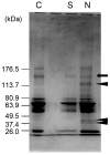
Protein adsorption profile on nano-fibrous and solid-walled scaffolds. Polyacrylamide gels stained with coomassie blue, lane C – bovine serum proteins, lane S – adsorbed bovine serum proteins on the solid-walled scaffold, lane N – adsorbed bovine serum proteins on the nano-fibrous scaffold. From Woo, Chen and Ma [99], Copyright © 2003 by John Wiley & Sons. Reprinted by permission of John Wiley & Sons.
Figure 5
Figure 5
Relative levels of bone marker gene expression on NF and SW scaffolds after 2 and 6 weeks of culture under differentiation conditions. (A) Osteocalcin (OCN) expression. (B) Bone sialoprotein (BSP) expression. From Chen, Smith and Ma [100], Copyright © 2006 by Elsevier.
Figure 6
Figure 6
Effect of dehydroproline, an inhibitor of collagen fiber formation, on gene expression in neonatal mouse calvarial osteoblasts grown on the scaffolds. From Woo, Jun, Chen, Seo, Baek, Ryoo, Kim, Somerman and Ma [103], Copyright © 2006 by Elsevier.
Figure 7
Figure 7
SEM micrographs of PLLA/mHAP (A, B) and PLLA/nHAP (C, D) composite scaffolds fabricated using phase separation. (A, B) From Zhang and Ma [11], Copyright © 1999 by John Wiley & Sons. Reprinted with permission of John Wiley & Sons; (C, D) From Wei and Ma [109], Copyright © 2004 by Elsevier.
Figure 8
Figure 8
SEM micrographs of PLLA/apatite composite scaffold prepared by a biomimetic process in a simulated body fluid (SBF). (A, B) PLLA scaffolds phase separated in dioxane; (C, D) PLLA nano-fibrous scaffolds prepared by sugar template leaching and phase separation in THF. Scaffolds were incubated in 1.5x SBF at 37 °C for 30 days. (A, B) From Zhang and Ma [82], Copyright © 1999 by John Wiley & Sons; (C, D) From Wei and Ma [94], Copyright © 2006 by John Wiley & Sons. Reprinted with permission of John Wiley & Sons.
Figure 9
Figure 9
Scanning electron micrographs of nanosphere (NS) incorporated PLLA nano-fibrous scaffolds: (A, B) PLLA nano-fibrous scaffolds before nanosphere incorporation at 100x (A) and 10,000x (B); (C, D) PLLA nano-fibrous scaffolds after nanosphere incorporation at 100x (C) and 10,000x (D). From Wei, Jin, Giannobile and Ma [140], Copyright © 2007 by Elsevier.
Figure 10
Figure 10
In vitro release kinetics of rhBMP-7 from nanospheres immobilized on nano-fibrous scaffolds: In 10 mM PBS with a BMP-7 loading of 200 ng/scaffold. Each data point represents a mean ± standard deviation (n=3). From Wei, Jin, Giannobile and Ma [140], Copyright © 2007 by Elsevier.
Figure 11
Figure 11
BMP7-NS incorporated nano-fibrous PLLA scaffolds were implanted subcutaneously onto the dorsa of rats for 6 weeks: (A) Significant bone formation in the BMP7-NS containing scaffolds (H & E staining); (B) Fibrous tissue in the scaffolds without BMP7 (H & E); (C) Fibrous tissue in scaffolds pre-soaked in a BMP7 solution (H & E). From Wei, Jin, Giannobile and Ma [140], Copyright © 2007 by Elsevier.

References

    1. Langer R, Vacanti JP. Tissue engineering. Science. 1993;260:920–6. - PubMed
    1. Ma PX. Scaffolds for tissue fabrication. Materials Today. 2004;7:30–40.
    1. Ma PX. Tissue Engineering. In: Kroschwitz JI, editor. Encyclopedia of Polymer Science and Technology. Third. Vol. 12. John Wiley & Sons, Inc.; Hoboken, NJ: 2005. pp. 261–291.
    1. Rice MA, Dodson BT, Arthur JA, Anseth KS. Cell-based therapies and tissue engineering. Otolaryngol Clin North Am. 2005;38:199–214. - PubMed
    1. Liu X, Ma PX. Polymeric scaffolds for bone tissue engineering. Annals of biomedical engineering. 2004;32:477–486. - PubMed

Publication types

MeSH terms

Substances