Case Reports
. 2007 Nov 27;104(48):19073-8.
doi: 10.1073/pnas.0704336104.

A T-cell receptor associated with naturally occurring human tumor immunity

Affiliations
Case Reports

A T-cell receptor associated with naturally occurring human tumor immunity

Bianca D Santomasso et al. Proc Natl Acad Sci U S A. .

Abstract

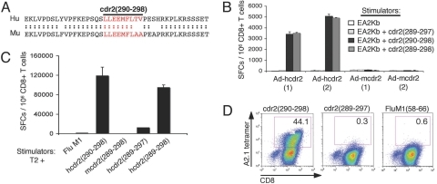
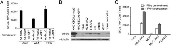
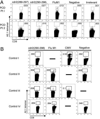
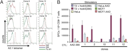
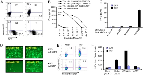
The onconeural antigens appear to serve as tumor rejection antigens in the paraneoplastic neurologic disorders. Here, we used an unbiased peptide binding screen, followed by studies in HLA-A2.1 transgenic mice to identify naturally processed HLA-A2.1 restricted epitopes of the paraneoplastic cerebellar degeneration breast/ovarian cancer antigen cdr2. These mice were used to clone high-avidity cdr2-specific CD8(+) T cells that recognize human tumor cells presenting endogenously loaded MHC class I-cdr2 peptide. T cells with this specificity were detected in the peripheral blood of two HLA-A2.1(+) paraneoplastic cerebellar degeneration patients. We cloned T cell receptor (TCR) alpha and beta genes from cdr2-specific T cells; electroporation of RNA encoding this TCR turned nonreactive donor T cells into efficient killers of human cdr2-expressing tumor cells. Cloned cdr2-specific TCR genes provide a clinically relevant means for immunologic targeting of human gynecologic cancers.


Conflict of interest statement

The authors declare no conflict of interest.

Figures

Fig. 1.
Fig. 1.
Characterization of cdr2 (290)-specific CTLs from Ad-hcdr2 immunized A2.1 transgenic mice. (A) Sequence homology of murine (Mu) and human (Hu) cdr2 surrounding residues 290–298. (B) AAD A2.1 transgenic mice were immunized with 109 pfu of Ad-hcdr2 or Ad-mcdr2 (two animals per group), and 12 days later splenic CD8+ T cells were cocultured with EA2Kb cells pulsed with human cdr2 (289–297), cdr2 (290), or cdr2 (289–298) peptide (10−5 M) in an 18-h IFN-γ ELISPOT assay. Spot-forming cells (SFCs) per 106 CD8+ T cells are shown (the average of triplicate wells, error bars indicate standard deviations). (C) Splenocytes from Ad-hcdr2-immunized AAD mice were stimulated with human cdr2 (290) for two rounds in vitro and assayed by IFN-γ ELISPOT using indicated peptide-pulsed T2 cells as stimulators. (D) CD8-independence of AAD 290 CTL. AAD 290 CTL were purified by CD8-negative selection and stained with the indicated HLA-A2.1 tetramers.
Fig. 2.
Fig. 2.
AAD 290 CTL recognize endogenous cdr2. (A) AAD 290 CTL were cocultured with KECs isolated from AAD, AAA, or HHD mice; KECs were infected with the indicated AdV constructs and used as stimulators in IFN-γ ELISPOT assay. (B) Western blot analysis of cdr2 protein expression tumor cell lines and normal tissue. The antibody used detects cdr2 and cdr3 as discussed (10); additional IP-Western blot analysis with a cdr2-specific antibody confirmed cdr2 expression from Ad-hcdr2, HeLa, COV413, and mouse cerebellum (data not shown). (C) AAD 290 CTL recognizes endogenous cdr2 in tumors. AAD 290 CTL were cocultured with the indicated tumor cells with (gray) or without (white) pretreatment with IFN-γ for 40 h before ELISPOT assay.
Fig. 3.
Fig. 3.
Tetramer analysis of human CD8+ T cells in HLA-A2.1+ PCD patients. (A) PBLs from two A2.1+ PCD patients were stained with tetramers specific to cdr2, FluM1 (58–66), control [PSMA (4–12) in Pt 1, HuD (157–165) in Pt 2], or negative (Beckman Coulter) tetramer and analyzed by flow cytometry. Results are gated on CD8+ T cells. (B) Tetramer analysis of control patients: normal donor (I), two Hu PND patients (II and III), and a neurologically normal ovarian cancer patient (IV). Controls are FluM1 (Pt 1 harbored FluM1 tetramer-positive CD8+ T cells confirmed by IFN-γ ELISPOT assay; Pt 2 had no flu response), CMVpp65 (495–503), or negative A2.1 tetramers.
Fig. 4.
Fig. 4.
Structural and functional avidity of cdr2-specific T cell clones. (A) CD8+ T cells from the AAD 290 CTL line (after the 6th in vitro restimulation) and eight daughter CTL clones were stained with A2.1/cdr2 (290) tetramer (red line) or control [A2.1/cdr2 (289–297) (blue line), or FluM1 (green line)] tetramers and analyzed by FACS. (B) The ability of bulk AAD 290 CTL or the indicated clones to recognize human cdr2 (289–297) or cdr2 (290) peptide-pulsed T2 cells (10−6 M) or the indicated cdr2-expressing tumor cells (pretreated with IFN-γ for 40 h) was evaluated in an 18-h IFN-γ ELIPSOT assay.
Fig. 5.
Fig. 5.
Characterization of a cdr2-specific T cell receptor. (A) Tetramer analysis of CD8+ PBL electroporated with cdr2-TCR mRNA. cdr2 TCR α- and β-chain (1 and 2) and GFP RNAs (3 and 4) were electroporated into 106 OKT3-stimulated CD8+ PBL. FACS analysis with negative (1 and 3) or cdr2 (290) tetramer (2 and 4) and percentage staining cells is shown. (B–F) Functional analysis of cdr2 TCR. Human CD8+ T cells were electroporated with cdr2 TCR, cocultured for 18 h with target cells [T2 cells pulsed with the indicated peptide (B), or AAA KECs-transduced with the indicated AdV constructs (C)], and IFN-γ secretion measured by ELISA. Values are the average of duplicates. In B, IFN-γ secretion in cocultures with GFP-electroporated CD8+ T cells was <15 pg/ml for all peptides (data not shown). (D) The cells in C were assessed for cell lysis by fluorescence microscopy. (E) FACS analysis for surface expression of CD107a after mock- or TCR-electroporated human CD8+ T cells were cocultured with Ad-hcdr2 or Ad-GFP-transduced AAA (A2.1) KECs. (F) cdr2 TCR-electroporated CD8+ T cells recognize cdr2-expressing gynecologic tumor cells. Human CD8+ T cells were electroporated with cdr2 TCR α- and β-chain (black bar) or GFP (blue bar) RNA and cocultured with the indicated tumor cells (5 × 10⁴) in an 18-h IFN-γ ELISPOT assay; average of duplicate wells and SD are shown.

References

    1. Rosenberg SA. Nature. 2001;411:380–384. - PubMed
    1. Dudley ME, Rosenberg SA. Nat Rev Cancer. 2003;3:666–675. - PMC - PubMed
    1. Boon T, Coulie PG, Van den Eynde BJ, van der Bruggen P. Annu Rev Immunol. 2006;24:175–208. - PubMed
    1. Darnell RB. Proc Natl Acad Sci USA. 1996;93:4529–4536. - PMC - PubMed
    1. Darnell RB, Posner JB. Nat Immunol. 2003;4:201. - PubMed

Publication types

MeSH terms