. 2008 Jan;135(1):43-52.
doi: 10.1242/dev.011288. Epub 2007 Nov 28.

The E1 ubiquitin-activating enzyme Uba1 in Drosophila controls apoptosis autonomously and tissue growth non-autonomously

Affiliations

The E1 ubiquitin-activating enzyme Uba1 in Drosophila controls apoptosis autonomously and tissue growth non-autonomously

Tom V Lee et al. Development. 2008 Jan.

Abstract

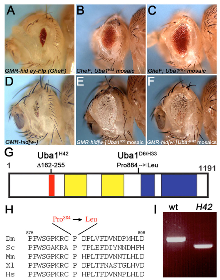
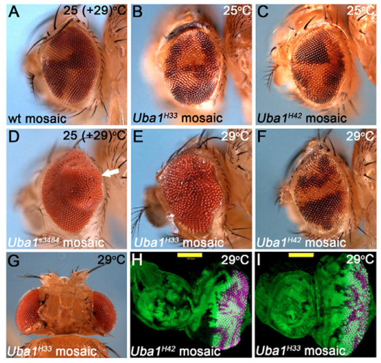
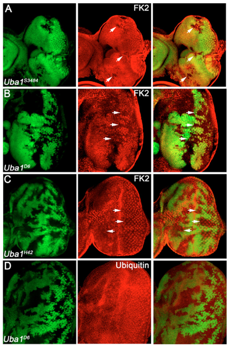
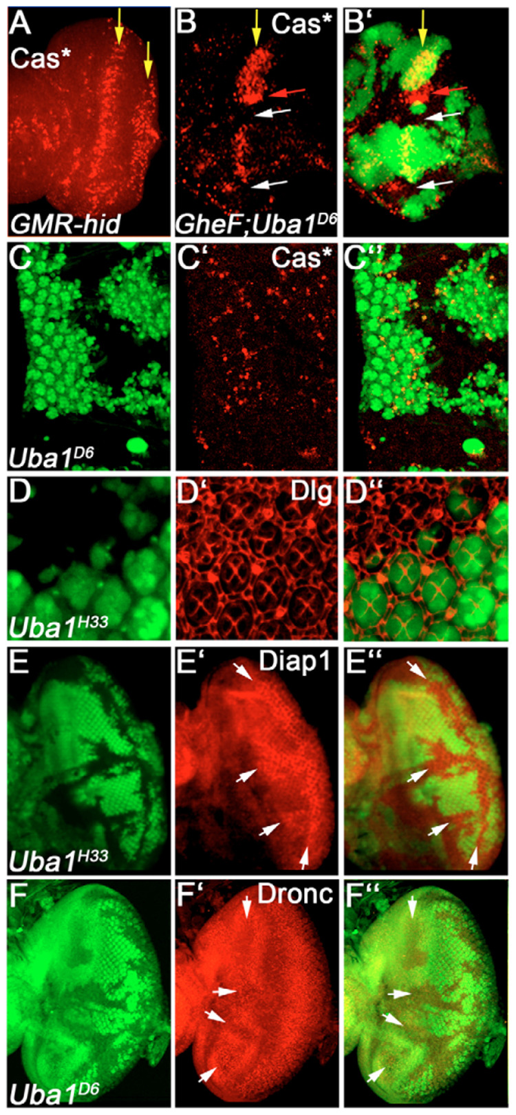
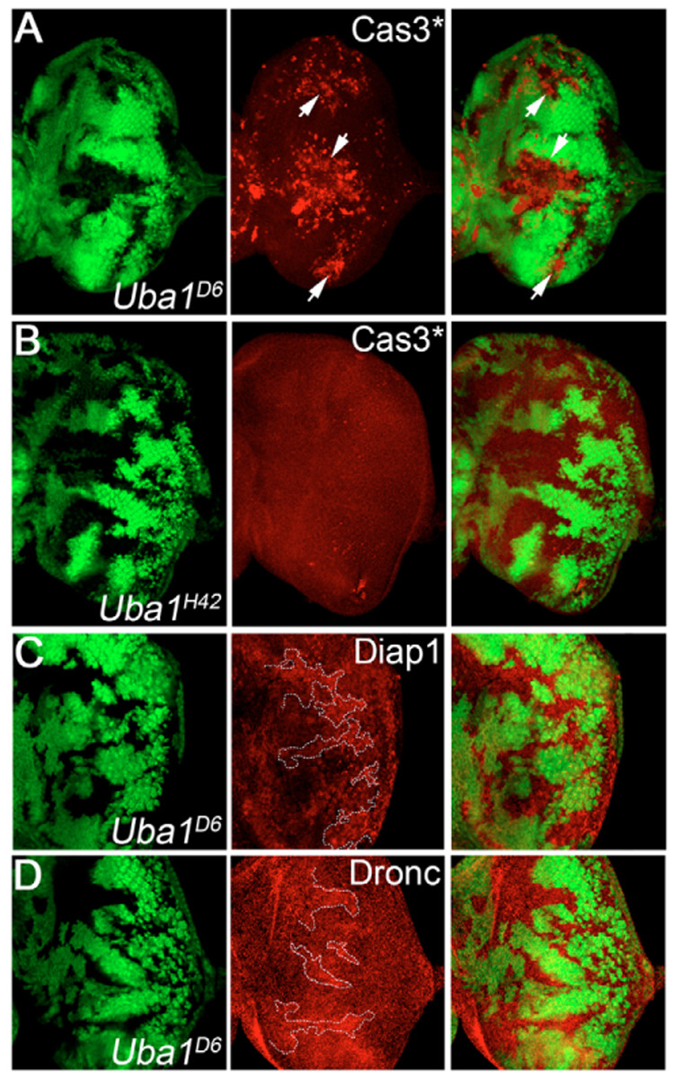
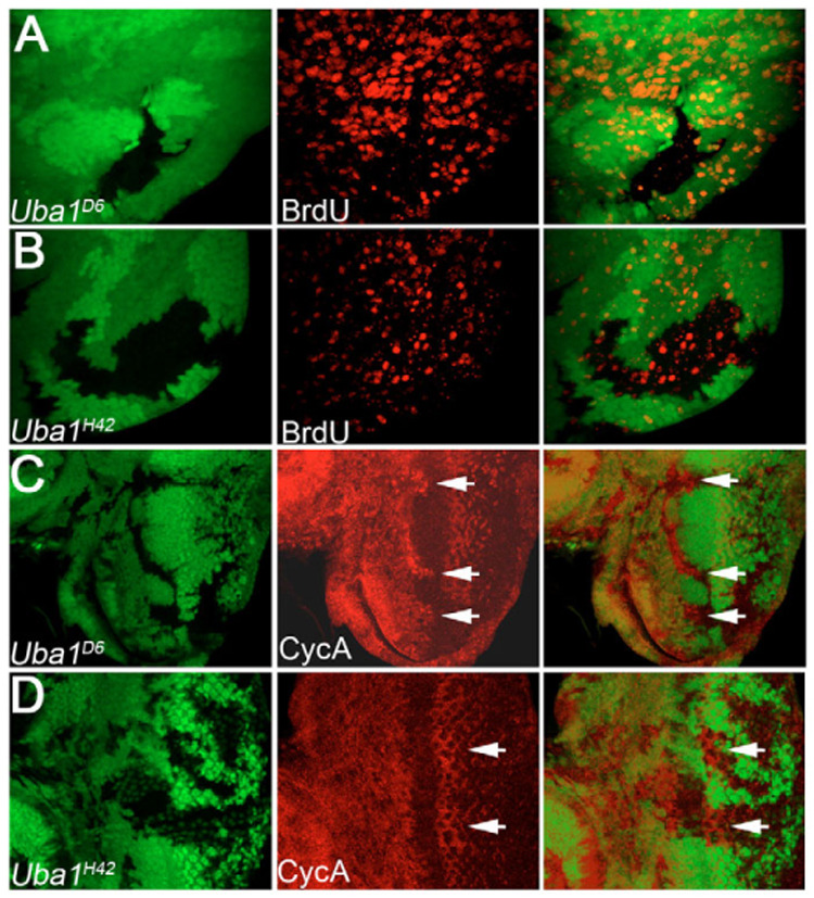
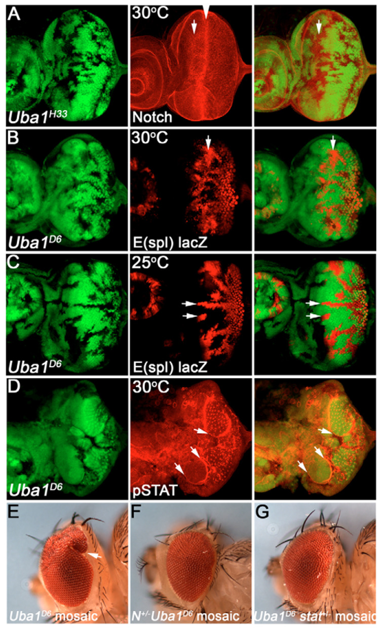
Ubiquitination is an essential process regulating turnover of proteins for basic cellular processes such as the cell cycle and cell death (apoptosis). Ubiquitination is initiated by ubiquitin-activating enzymes (E1), which activate and transfer ubiquitin to ubiquitin-conjugating enzymes (E2). Conjugation of target proteins with ubiquitin is then mediated by ubiquitin ligases (E3). Ubiquitination has been well characterized using mammalian cell lines and yeast genetics. However, the consequences of partial or complete loss of ubiquitin conjugation in a multi-cellular organism are not well understood. Here, we report the characterization of Uba1, the only E1 in Drosophila. We found that weak and strong Uba1 alleles behave genetically differently with sometimes opposing phenotypes. Whereas weak Uba1 alleles protect cells from cell death, clones of strong Uba1 alleles are highly apoptotic. Strong Uba1 alleles cause cell cycle arrest which correlates with failure to reduce cyclin levels. Surprisingly, clones of strong Uba1 mutants stimulate neighboring wild-type tissue to undergo cell division in a non-autonomous manner giving rise to overgrowth phenotypes of the mosaic fly. We demonstrate that the non-autonomous overgrowth is caused by failure to downregulate Notch signaling in Uba1 mutant clones. In summary, the phenotypic analysis of Uba1 demonstrates that impaired ubiquitin conjugation has significant consequences for the organism, and may implicate Uba1 as a tumor suppressor gene.


Figures

Fig. 1
Fig. 1. Uba1 mutants are autonomous suppressors of GMR-hid-induced apoptosis
(A) The unmodified small eye phenotype caused by GMR-hid ey-FLP (GheF). (B,C) The GheF phenotype is moderately strongly suppressed by Uba1 alleles at 25°C. Genotype: GheF; FRT42D Uba1H33 and Uba1H42/FRT42 P[w+], respectively. (D) The unmodified small eye phenotype caused by a GMR-hid transgene that lacks the white+ marker gene (GMR-hid[w]). Note, the GMR-hid transgene in (A) carries the white+ minigene. Genotype: ey-FLP; GMR-hid[w]. (E,F) The suppression of GMR-hid[w] is mediated by Uba1 mutant tissue which is phenotypically white. Flies were incubated at 25°C. Genotype: ey-FLP; FRT42D Uba1H33 and Uba1H42/FRT42D P[w+]; GMR-hid[w], respectively. (G) Outline of the Uba1 protein and locations of the mutations of the Uba1 alleles isolated in this study. The red box indicates the internal deletion of Uba1H42. The yellow boxes mark the Thif domains, and the blue boxes highlight the location of the catalytic Ube domains. (H) A highly conserved stretch of residues, incorporating Pro884 is present in range of species from yeast to humans. Dm, Drosophila melanogaster; Sc, Saccharomyces cerevisiae; Mm, Mus musculus; XI, Xenopus laevis; Hs, Homo sapiens. (I) RT-PCR of mRNA isolated from wild-type and Uba1H42 mutant larvae revealing an aberrantly spliced product. This results in an in frame deletion from amino acid residue 162 to 255 of Uba1H42 (see G). Primers were directed against the N-terminal third of Uba1.
Fig. 2
Fig. 2. Strong Uba1 alleles cause non-autonomous overgrowth
(A) Control mosaics: ey-FLP-induced clones in wild-type background. This phenotype is identical at 25°C and 29°C (shown is 25°C). Genotype: ey-FLP; FRT42D+/FRT42D P[w+]. (B,C) Clones of weak Uba1 mutants (phenotypically white) develop normally. Genotype: ey-FLP; FRT42D Uba1H33 and Uba1H42/FRT42D P[w+], respectively. (D) Eye mosaics of the strong Uba1s3484 allele are overgrown with bulges (arrow). Genotype: ey-FLP;FRT42D Uba1s3484/FRT42D P[w+]. This phenotype was obtained at 25°C, and is similar at 29°C. (E) Eye mosaic of the ts allele Uba1H33 at 29°C. Uba1H33 clones, which would be phenotypically white, are not detectable. The eyes appear rough, irregular and are often enlarged compared to wild-type eyes. Identical results were obtained for Uba1D6. Genotype as in B. (F) Eye mosaic of the weak Uba1H42 allele at 29°C. This allele does not behave as a ts allele. Phenotype and genotype are identical to those at 25°C (C). (G) Mosaic head of an Uba1D6 animal raised at 29°C. The left half of the Uba1H33 mosaic head is larger than the right half, suggesting that the overgrowth phenotype depends on the number and location of the Uba1 clones. Genotype as in B. (H,I) Eye-antennal imaginal discs of wild-type (H) and Uba1D6 (I) mosaic animals raised at 29°C. These discs were labeled with Elav antibody which labels photoreceptor neurons to identify discs of comparable age. Photoreceptor differentiation does occur in strong Uba1 clones, but is weakly delayed (see also Fig. S1 in the supplementary material). Genotype as in A and B, respectively. Size bar: 100 µm.
Fig. 3
Fig. 3. Uba1 mutant clones affect ubiquitin conjugation
(A–D) The left panels indicate the positions of the Uba1 clones by absence of GFP, the middle panels show the experiment and the right panels are the merged images of left and middle panels. Genotype in all panels: ey-FLP; FRT42D Uba1/FRT42D P[ubi-GFP]. The allele is indicated in the panels. (A–C) Uba1 mutant discs were labeled with the FK2 antibody which specifically recognizes mono- and poly-ubiquitinated conjugates, but not unconjugated ubiquitin (Fujimuro et al., 1994). The strong alleles Uba1s3484 (A) and Uba1D6 (B, incubated at 29°C) significantly reduce FK2 immunoreactivity (see arrows). Clones of the weak allele Uba1H42 (C) mildly reduce FK2 immunoreactivity (arrows). (D) Uba1D6 mosaics incubated at 29°C. The overall protein levels of ubiquitin are unchanged in Uba1 mutant clones.
Fig. 4
Fig. 4. Weak Uba1 alleles suppress apoptosis
(A) GMR-hid eye disc labeled with cleaved Caspase-3 (Cas3*) antibodies. Yellow arrows indicate two zones of dying cells in GMR-hid. (B,B′) Suppression of Cas3*-positive cell death in GMR-hid discs. The yellow arrow indicates the position of the first zone of Cas3*-positive cell death in GMR-hid. Uba1D6 clones suppress Cas3*-positive cell death (white arrows). However, this suppression is not uniform and occurs in a region-specific manner. Clones in an area of the disc marked by the red arrow are still apoptotic. Genotype: GMR-hid ey-FLP; FRT42D Uba1D6/FRT42D P[ubi-GFP]; (C–C″) Uba1D6 clones suppress most normal developmental cell death in 28 hours APF pupal discs. Genotype as in B. (D–D″) Uba1H33 clones contain additional IOCs in 42 hours APF pupal discs. Genotype: ey-FLP; FRT42D Uba1H33/FRT42D P[ubi-GFP]. (E–E″) Diap1 protein levels are increased in Uba1H33 mutant clones (arrows). Genotype as in D. (F–F″) Dronc protein levels are decreased in Uba1D6 mutant clones (arrows). Genotype as in B.
Fig. 5
Fig. 5. The apoptotic phenotype of strong Uba1 alleles
All discs were incubated at 29°C. (A,C,D) ey-FLP; FRT Uba1D6/FRT P[ubi-GFP]; (B) ey-FLP; FRT Uba1H42/FRT P[ubi-GFP]. (A) Uba1D6 clones are highly Cas3*-apoptotic (see arrows). (B) Uba1H42 clones lack apoptotic labeling. (C) Uba1D6 clones (outlined by dashed line) contain increased levels of Diap1 protein. (D) Uba1D6 clones (outlined by dashed line) contain increased levels of Dronc protein.
Fig. 6
Fig. 6. Cell cycle arrest and non-autonomous proliferation in Uba1 mosaics
(A,B) Third instar mosaic Uba1D6 (A) and Uba1H42 (B) wing discs incubated at 29°C. Note lack of BrdU labeling in Uba1D6 mutant clones, and increased BrdU labeling in wild-type tissue surrounding Uba1D6 clones. Proliferation in Uba1H42 mosaics is homogenous within and outside the clones (B). (C,D) Cyclin A labeling of eye discs from third instar larval eye discs containing Uba1D6 (C) and Uba1H42 (D) mutant clones. Cyclin A levels are increased in Uba1D6 clones spanning the morphogenetic furrow (arrows in C). Clones of the weak allele Uba1H42 contain slightly increased Cyclin A levels (D, arrows). However, this increase does not affect cell proliferation (B).
Fig. 7
Fig. 7. Notch signaling induces non-autonomous overgrowth in Uba1 mosaics
All discs were incubated at 30°C, except C which was incubated at 25°C. (A) Uba1H33 mosaic eye discs labeled with Notch antibody C17.9C6 which was raised against the intracellular domain of Notch. Arrows indicate increase of Notch protein in Uba1 clones. This is most prominent immediately anterior to the morphogenetic furrow (arrowhead). Genotype: ey-FLP; FRT Uba1D6/FRT P[ubi-GFP]. (B,C) Notch activity as visualized by the Notch reporter E(spl)m8 2.61-lacZ is increased in Uba1D6 clones at 30°C (B) and 25°C (C). Genotype: ey-FLP; E(spl)m8 2.61-lacZ FRT ubaD6/FRT P[ubi-GFP]. (D) pSTAT labeling is increased in a non-autonomous manner in wild-type tissue immediately adjacent to Uba1 clones (arrow). Genotype as in A. (E) Eyes of Uba1D6 mosaic animals are overgrown with bulges (arrow). Genotype as in A. (F,G) Heterozygosity of Notch (F) and stat92 (G) dominantly suppresses the Uba1D6 overgrowth phenotype. Genotype: Notch264-39/ey-FLP; FRT42 Uba1D6/FRT42 P[ubi-GFP] (F) and ey-FLP; FRT42 Uba1D6/FRT42 P[ubi-GFP]; stat9285c9 (G).

References

    1. Babst M. A protein's final ESCRT. Traffic. 2005;6:2–9. - PubMed
    1. Cagan RL, Ready DF. The emergence of order in the Drosophila pupal retina. Dev. Biol. 1989;136:346–362. - PubMed
    1. Cashio P, Lee TV, Bergmann A. Genetic control of programmed cell death in Drosophila melanogaster. Semin. Cell Dev. Biol. 2005;16:225–235. - PubMed
    1. Chai J, Yan N, Huh JR, Wu JW, Li W, Hay BA, Shi Y. Molecular mechanism of Reaper-Grim-Hid-mediated suppression of DIAP1-dependent Dronc ubiquitination. Nat. Struct. Biol. 2003;10:892–898. - PubMed
    1. Chao JL, Tsai YC, Chiu SJ, Sun YH. Localized Notch signal acts through eyg and upd to promote global growth in Drosophila eye. Development. 2004;131:3839–3847. - PubMed

Publication types

MeSH terms