. 2008 Feb 8;283(6):3655-3664.
doi: 10.1074/jbc.M708375200. Epub 2007 Nov 28.

Structural and functional analyses of the severe acute respiratory syndrome coronavirus endoribonuclease Nsp15

Affiliations

Structural and functional analyses of the severe acute respiratory syndrome coronavirus endoribonuclease Nsp15

Kanchan Bhardwaj et al. J Biol Chem. .

Abstract

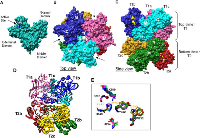
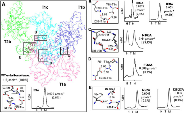
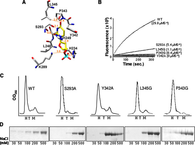
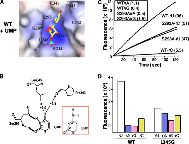
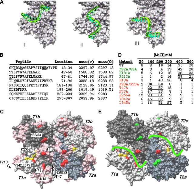
The severe acute respiratory syndrome (SARS) coronavirus encodes several RNA-processing enzymes that are unusual for RNA viruses, including Nsp15 (nonstructural protein 15), a hexameric endoribonuclease that preferentially cleaves 3' of uridines. We solved the structure of a catalytically inactive mutant version of Nsp15, which was crystallized as a hexamer. The structure contains unreported flexibility in the active site of each subunit. Substitutions in the active site residues serine 293 and proline 343 allowed Nsp15 to cleave at cytidylate, whereas mutation of leucine 345 rendered Nsp15 able to cleave at purines as well as pyrimidines. Mutations that targeted the residues involved in subunit interactions generally resulted in the formation of catalytically inactive monomers. The RNA-binding residues were mapped by a method linking reversible cross-linking, RNA affinity purification, and peptide fingerprinting. Alanine substitution of several residues in the RNA-contacting portion of Nsp15 did not affect hexamer formation but decreased the affinity of RNA binding and reduced endonuclease activity. This suggests a model for Nsp15 hexamer interaction with RNA.


Figures

FIGURE 1
FIGURE 1
Structure of SARS-CoV Nsp15.A, surface representation of a subunit showing three domains. Active site residues are colored by element and indicated by an arrow. B, arrangement of six subunits from top view of hexamer. N-terminal, middle, and C-terminal domains are indicated as N, M, and C, respectively. The arrows indicate the positions of catalytic sites in top trimer. C, side view of hexamer showing arrangement and N- to N-terminal interaction of the top trimer (T1) with bottom trimer (T2). Six subunits are labeled as a-c and are colored as follows: T1a, pink; T1b, blue; T1c, cyan; T2a, red; T2b, green; T2c, golden. This color scheme is used throughout the figures. Catalytic residues are colored yellow. D, worm diagram drawn based on b-factor. Worm thickness is directly proportional to flexibility, i.e. the thickest region indicates most flexible. E, overlap of the catalytic residues within the active sites of the six subunits. The structures are anchored by the backbond of His249.
FIGURE 2
FIGURE 2
Subunit interaction.A, wire diagram of subunit T1a and its interaction with other subunits. Contact regions are boxed and labeled (boxes B-E). The inset shows molecular arrangement and atomic distances between E3 and L2 (left) and the gel filtration profile of the E3A mutant of Nsp15 (20). H, T, and M denote positions corresponding to elution volume of a hexamer, trimer, and monomer of Nsp15, respectively. B-E, contacting residues and the calculated distances between them (left). Atomic distances are calculated using Chimera. Gel filtration chromatography elution profiles of the indicated mutant proteins (right). The values below the mutant name refer to the cleavage rate relative to WT in parentheses.
FIGURE 3
FIGURE 3
Analysis of the Nsp15 catalytic site.A, molecular docking of UMP into the Nsp15 active site. The backbone of active site residues is shown in gray, and that of UTP is in yellow; nitrogen and oxygen atoms are blue and red, respectively. The phosphorus is cyan. Atomic interaction is denoted by broken lines, and the calculated distances are indicated. B, results of cleavage of substrate rU with WT and mutant Nsp15, using real time fluorescent endoribonuclease assay. The identity of protein and rate of reaction is indicated to the right of the graph. The endoribonuclease activity was tested at 0.05 μm of WT or mutant proteins. C, Gel filtration of Nsp15. Elution profiles for WT Nsp15 and mutant versions as indicated next to the curve. The expected positions for hexamer (H), trimer (T), and monomer (M) are shown in each panel. D, SDS-PAGE analysis of polyU-agarose affinity chromatography fractions. WT or mutant Nsp15 proteins were incubated with polyU-agarose for 90 min at 30 mm NaCl. Unbound fraction was removed by a low speed centrifugation, and the slurry was washed sequentially with increasing NaCl concentration. Samples of each NaCl concentration wash were analyzed by SDS-PAGE and Coomassie Blue staining.
FIGURE 4
FIGURE 4
Examination of substrate specificity.A, UMP docked into catalytic pocket of WT Nsp15. B, schematic to denote uridine recognition by S293 and the approximate distances between UMP and nearby catalytic site residues. C, results of real time fluorescent endoribonuclease assay. 0.25 μm of WT or mutant S293A proteins were used in these assays, and cleavage of substrates rU, rC, rA, and rG was measured in real time. The results of rU and rC cleavage are shown in real time. The slopes of each curve represent the rate of reaction. The identities of protein and substrate along with rate of reaction are indicated to the right of the curves. Cleavage rates of rA and rG are shown in the inset. D, cleavage of fluorescent substrates by WT or L345G proteins. Each protein (0.25 μm) was incubated with one of the four fluorogenic substrates (rU, rC, rA, or rG) for 20 min at room temperature and measured the fluorescence for each treatment. The increased cleavage of the rA substrate by L345G was confirmed in kinetic assay in two other independent experiments (data not shown).
FIGURE 5
FIGURE 5
A Model for a complex between Nsp15 and oligonucleotide RNA.A, putative RNA binding path of sNsp15 hexamer. For clarity, only subunit T1A is emphasized. A U16 A-RNA oligonucleotide is manually docked through the narrow catalytic site of the enzyme. B, summary of Nsp15 peptides obtained by biotin-streptavidin affinity chromatography and reversible cross-linking and trypsin digestion. The peptides were purified using Zip-tip as recommended by the manufacturer and subjected to MALDI-ToF. The expected masses obtained by virtual tryptic digestion of Nsp15 are denoted by (e) and the masses of observed peptide peaks from MALDI-ToF analysis are denoted by (0). The peptide position on the protein sequence is indicated by protein sequence as well as residue numbers. C, location of peptides identified by reversible RNA cross-linking and MALDI-ToF analysis. Identified peptides are colored pink. Potential RNA binding residues, along trajectories selected for mutational analysis are colored red. Active site residues are colored by element. D, affinity chromatography analysis of Nsp15 mutant and wild type proteins. All of the proteins were allowed to bind to polyU-agarose at 30 mm sodium chloride concentration followed by washing the slurry with sodium chloride concentrations indicated on top. Protein in each fraction was quantified by the Coomassie Plus protein assay kit from Pierce and is indicated as a percentage of the amount loaded. E, favored RNA binding path on Nsp15. Color scheme is as follows: protein subunits, shades of gray; active site residues, yellow; residues that affected affinity of protein for RNA, red; residues that did not affect RNA binding, green. A green line indicating RNA path is drawn on protein surface.

References

    1. Gorbalenya A.E., Enjuanes L., Ziebuhr J., Snijder E.J. Virus Res. 2006;117:17–37. - PMC - PubMed
    1. Lai M.M., Cavanagh D. Adv. Virus Res. 1997;48:1–100. - PMC - PubMed
    1. Spaan W., Cavanagh D., Horzinek M.C. J. Gen. Virol. 1988;69:2939–2952. - PubMed
    1. Kim Y.C., Russell W.K., Ranjith-Kumar C.T., Thomson M., Russell D.H., Kao C.C. J. Biol. Chem. 2005;280:38011–38019. - PubMed
    1. Sawicki S.G., Sawicki D.L. Adv. Exp. Med. Biol. 1995;380:499–506. - PubMed

Publication types

MeSH terms

Associated data

LinkOut - more resources