. 2007 Nov 28;27(48):13065-73.
doi: 10.1523/JNEUROSCI.3467-07.2007.

Bradykinin-induced microglial migration mediated by B1-bradykinin receptors depends on Ca2+ influx via reverse-mode activity of the Na+/Ca2+ exchanger

Affiliations

Bradykinin-induced microglial migration mediated by B1-bradykinin receptors depends on Ca2+ influx via reverse-mode activity of the Na+/Ca2+ exchanger

Masataka Ifuku et al. J Neurosci. .

Abstract

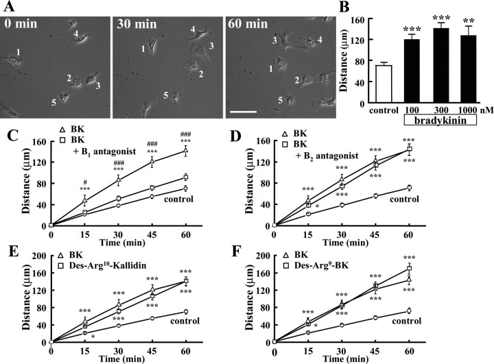
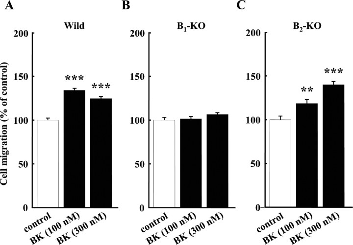
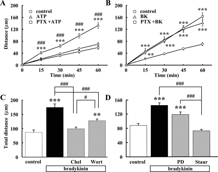
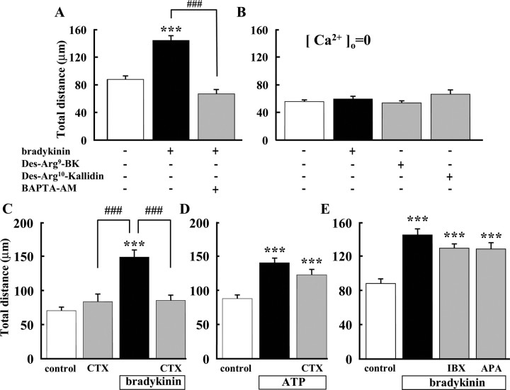
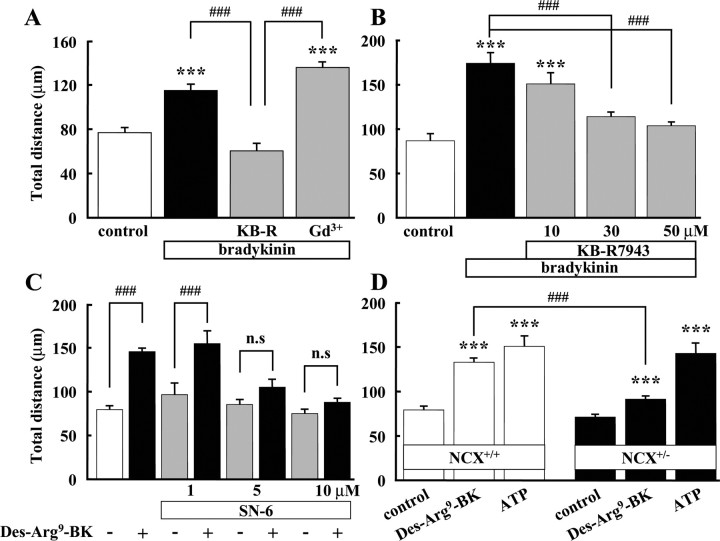
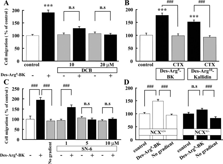
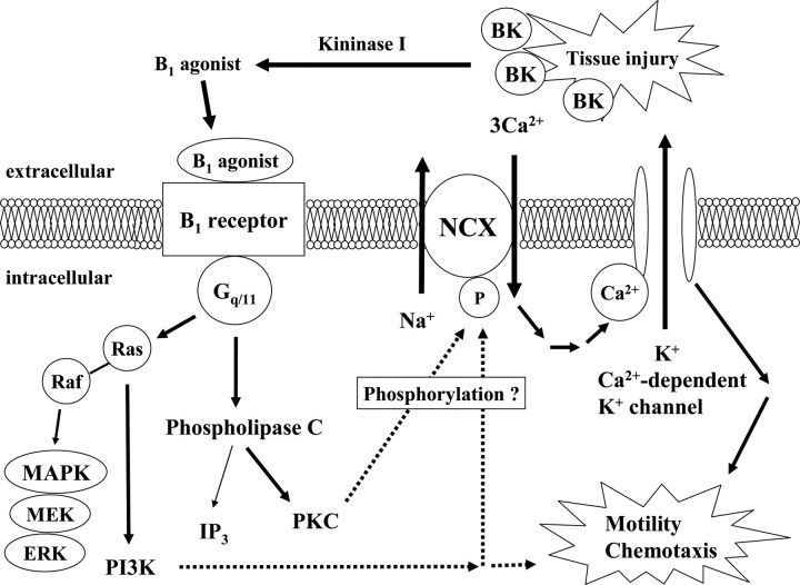
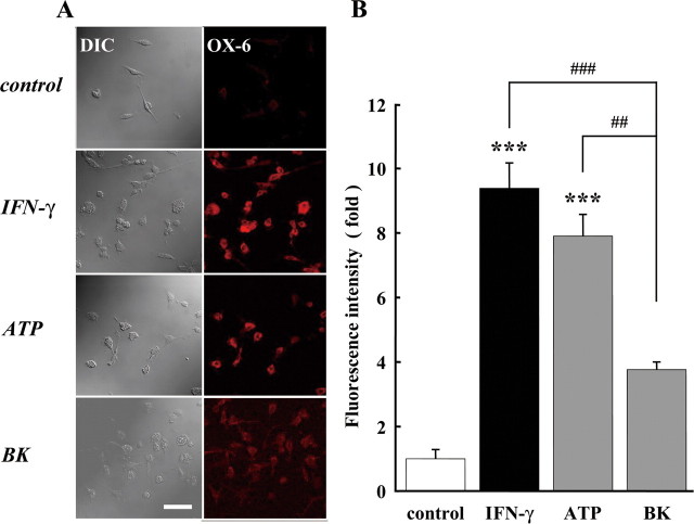
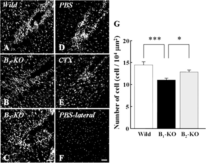
Bradykinin (BK) is produced and acts at the site of injury and inflammation. In the CNS, migration of microglia toward the lesion site plays an important role pathologically. In the present study, we investigated the effect of BK on microglial migration. Increased motility of cultured microglia was mimicked by B1 receptor agonists and markedly inhibited by a B1 antagonist but not by a B2 receptor antagonist. BK induced chemotaxis in microglia isolated from wild-type and B2-knock-out mice but not from B1-knock-out mice. BK-induced motility was not blocked by pertussis toxin but was blocked by chelating intracellular Ca2+ or by low extracellular Ca2+, implying that Ca2+ influx is prerequisite. Blocking the reverse mode of Na+/Ca2+ exchanger (NCX) completely inhibited BK-induced migration. The involvement of NCX was further confirmed by using NCX+/- mice; B1-agonist-induced motility and chemotaxis was decreased compared with that in NCX+/+ mice. Activation of NCX seemed to be dependent on protein kinase C and phosphoinositide 3-kinase, and resultant activation of intermediate-conductance (IK-type) Ca2+-dependent K+ currents (I(K(Ca))) was activated. Despite these effects, BK did not activate microglia, as judged from OX6 staining. Using in vivo lesion models and pharmacological injection to the brain, it was shown that microglial accumulation around the lesion was also dependent on B1 receptors and I(K(Ca)). These observations support the view that BK functions as a chemoattractant by using the distinct signal pathways in the brain and, thus, attracts microglia to the lesion site in vivo.


Figures

Figure 1.
Figure 1.
Motility of BK-stimulated microglial cells. A, Microglial cells were stimulated with 300 nm BK and were recorded using time-laps imaging system every 5 min. The images show at 0, 30, and 60 min after application of BK. B, Effects of different concentration of BK on microglial motility. Total distance (in micrometers) of microglial cells for 1 h was significantly increased by BK at 100, 300, and 1000 nm. **p < 0.01; ***p < 0.001. Effects of bradykinin receptor antagonist and agonist. C, B1 receptor antagonist, Des-Arg9-[Leu8]-bradykinin (1 μm), inhibited BK-induced microglial motility. ***p < 0.001, significantly different from control. #p < 0.05, ###p < 0.001, significantly different from B1 antagonist. D, B2 receptor antagonist, Hoe140 (1 μm), did not inhibit BK-induced microglial motility. ***p < 0.001. E, Total distance (in micrometers) of microglial migration for 1 h was increased by B1 agonist, Des-Arg10-kallidin (300 nm). F, Another B1 agonist, Des-Arg9-bradykinin (300 nm), also increased microglial motility. *p < 0.05; ***p < 0.001.
Figure 2.
Figure 2.
Microglial chemotaxis in three genotypes of bradykinin receptors. A, BK-induced microglial chemotaxis in wild-type (Wild) mice. B, In B1 receptor-knock-out mice (B1-KO), 100 and 300 nm BK did not induce chemotaxis. C, In B2 receptor knock-out mice (B2-KO), BK at either concentration induced microglial chemotaxis. **p < 0.005; ***p < 0.001.
Figure 3.
Figure 3.
Effects of PTX and inhibitors of protein kinases. A, Total distance (in micrometers) of microglial migration for 1 h of microglial cells was increased by ATP. Pretreatment of PTX (50 ng/ml) for 12 h (with additional 50 ng/ml PTX before application of ATP) almost completely blocked the effect of ATP. ***p < 0.001, significantly different from control. ###p < 0.001, significantly different from ATP+PTX. B, PTX did not affect BK (300 nm)-induced microglial motility (n = 20). ***p < 0.001. C, BK-induced microglial motility was inhibited by pretreatment of PKC inhibitor, chelerythrine (Chel; 10 μm), for 24 h. A part of BK-induced microglial motility was inhibited by pretreatment of PI3 kinase inhibitor, wortmannin (Wort; 1 μm), for 24 h. ***p < 0.001; ###p < 0.001. D, BK-induced microglial motility was inhibited by pretreatment of PKC inhibitor, staurosporine (Staur; 10 nm) for 24 h. However, BK-induced microglial motility was not affected by pretreatment of MAP/ERK inhibitor, PD98059 (PD; 10 μm), 24 h. ***p < 0.001; ###p < 0.001.
Figure 4.
Figure 4.
Extracellular Ca2+ and activation of intermediate-conductance IK(Ca) was important for BK-induced microglial motility. A, Microglial motility in the presence of BK was significantly inhibited by pretreatment with BAPTA-AM (10 μm) for 20 min. ***p < 0.001; ###p < 0.001. B, In the absence of extracellular Ca2+ ([Ca2+]o = 0), total distance (in micrometers) of microglial migration was not affected by BK (300 nm) (n = 19), Des-Arg9-bradykinin (300 nm) (n = 18), or Des-Arg10-kallidin (300 nm) (n = 18). C, BK-induced increase in microglial motility was inhibited by CTX (0.1 μm). D, ATP (50 μm)-induced microglial motility was not affected by CTX (0.1 μm). E, Iberiotoxin (IBX) and apamin (APA), LK-type (large-conductance) and SK-type (small-conductance) IK(Ca) blockers, respectively, had no effect on BK-induced microglial motility. ***p < 0.001; ###p < 0.001.
Figure 5.
Figure 5.
Activation of reverse mode of Na+/Ca2+ exchanger was important for bradykinin-induced microglial motility. A, BK-induced microglial motility was inhibited by KB-R7943, a specific inhibitor of reverse-mode NCX. BK-induced microglial motility was not affected by gadolinium (Gd3+, 100 μm), an inhibitor of capacitative Ca2+ channels. ***p < 0.001; ###p < 0.001. B, BK-induced microglial motility was inhibited by KB-R7943 dose dependently. ***p < 0.001; ###p < 0.001. C, B1 agonist (Des-Arg9-BK)-induced microglial motility was inhibited by SN-6 (5 and 10 μm), a more specific inhibitor of reverse-mode NCX. D, B1 agonist (Des-Arg9-BK)-induced microglial motility in wild-type (NCX+/+) mice (open bar) and NCX knock-out (NCX+/−) mice (filled bar). In NCX+/+ mice, 300 nm Des-Arg9-BK induced microglial motility was 1.7-fold increased, whereas in NCX+/− mice, Des-Arg9-BK-induced microglial motility was only 1.3-fold increased. ***p < 0.001; ###p < 0.001. n.s., No significant difference.
Figure 6.
Figure 6.
B1 agonist-induced microglial chemotaxis, determined by Boyden chamber, was dependent on NCX and IK(Ca) activity. A, Des-Arg9-bradykinin (300 nm)-induced microglial chemotaxis was inhibited by DCB (10 and 20 μm), an inhibitor of NCX. ***p < 0.001; ###p < 0.001. B, CTX (0.1 μm) inhibited microglial chemotaxis induced by B1 agonists, Des-Arg9-bradykinin (300 nm) and Des-Arg10-kallidin (300 nm). ***p < 0.001; ###p < 0.001. C, Des-Arg9-bradykinin (300 nm)-induced microglial chemotaxis was inhibited by SN-6 (5 and 10 μm). ***p < 0.001; ###p < 0.001. D, Des-Arg9-bradykinin (300 nm) induced microglial chemotaxis in wild-type (NCX+/+) mice (open bar), but not in NCX knock-out (NCX+/−) mice or when Des-Arg9-BK was added to both upper and lower wells to eliminate the concentration gradient (No gradient). ###p < 0.001. n.s., No significant difference.
Figure 7.
Figure 7.
Possible signal transduction caused by activation of B1 receptor. Bradykinin is produced at site of tissue injury. Bradykinin is rapidly degraded by peptidases known as kininase I, which cleave off C-terminal arginine (Arg9) to generate Des-Arg9-bradykinin. Activation of the B1 receptor and coupling to Gq/11 results in the activation of PLCβ and PKC. PKC can phosphorylate NCX and increase NCX activity and subsequent mobilization of intracellular Ca2+. Increase in intercellular Ca2+ can activate Ca2+-dependent K+ channels. Resulting hyperpolarization may induce microglial motility and chemotaxis. Activation of the B1 agonist also couples the B1 receptor to MAPK signaling, which involves activation of Ras and consecutive stimulation of PI3K. PI3K was involved in a part of microglial motility.
Figure 8.
Figure 8.
Bradykinin did not stimulate MHCII expression. A, The cells were treated with 50 U/ml IFN-γ, 50 μm ATP, or 300 nm BK for 24 h. Differential interference contrast (DIC) image and immunofluorescence stained with anti-OX6 antibody (labeled with Alexa Fluor 568; red) are shown. Scale, 100 μm. B, The relative fluorescence intensity of OX6 immunoreactivity. The intensity of fluorescence after application of IFN-γ, ATP, or BK stimulation was normalized to the one in untreated (control) microglia. ***p < 0.001; ##p < 0.01; ###p < 0.001.
Figure 9.
Figure 9.
Bradykinin acts as an inducer of microglial migration in vivo. A–C, B1 receptors play an important role in migration and accumulation of microglia. Microglial cells around lesion were stained immunohistochemically with anti-Iba-1 in wild-type (A), B1-knock-out (B), and B2-knock-out mice (C). D, E, Ca2+-dependent K+ channels play an important role in migration and accumulation of microglia. Injection of CTX inhibited the number of microglia around lesion (E) compared with that in control (PBS-injected; D). F, Microglial distribution at contralateral cortex in control brain (D). G, The number of microglia around lesion was decreased in B1-knock-out mice and CTX-injected brain compared with the one in wild-type and B2-knock-out mice and control (PBS-injected) brain, respectively. Numbers of cells are represented as mean ± SEM. *p < 0.05; ***p < 0.001. Scale bar, 100 μm.

Comment in

  • Bradykinin: a microglia attractant in vivo?
    Huisman C, Kok P, Schmaal L, Verhoog P. Huisman C, et al. J Neurosci. 2008 Apr 2;28(14):3531-2. doi: 10.1523/JNEUROSCI.0374-08.2008. J Neurosci. 2008. PMID: 18385310 Free PMC article. No abstract available.

Similar articles

Cited by

References

    1. Abbott NJ. Inflammatory mediators and modulation of blood-brain barrier permeability. Cell Mol Neurobiol. 2000;20:131–147. - PMC - PubMed
    1. Busser BW, Hammond TG, Bjorling DE, Saban R. Lipopolysaccharide upregulates bradykinin 1 receptors in the isolated mouse bladder. J Urol. 1998;160:2267–2273. - PubMed
    1. Campos MM, Souza GE, Calixto JB. Upregulation of B1 receptor mediating des-Arg9-BK-induced rat paw oedema by systemic treatment with bacterial endotoxin. Br J Pharmacol. 1996;117:793–798. - PMC - PubMed
    1. Davalos D, Grutzendler J, Yang G, Kim JV, Zuo Y, Jung S, Littman DR, Dustin ML, Gan WB. ATP mediates rapid microglial response to local brain injury in vivo. Nat Neurosci. 2005;8:752–758. - PubMed
    1. Delmas P, Wananerbercq N, Abogadie FC, Mistry M, Brown DA. Signaling microdomains define the specificity of receptor-mediated InsP3 pathways in neurons. Neuron. 2002;14:209–220. - PubMed

Publication types

MeSH terms

LinkOut - more resources