. 2007 Nov 28;27(48):13117-29.
doi: 10.1523/JNEUROSCI.3322-07.2007.

Cdc42 regulates cofilin during the establishment of neuronal polarity

Affiliations

Cdc42 regulates cofilin during the establishment of neuronal polarity

Boyan K Garvalov et al. J Neurosci. .

Abstract

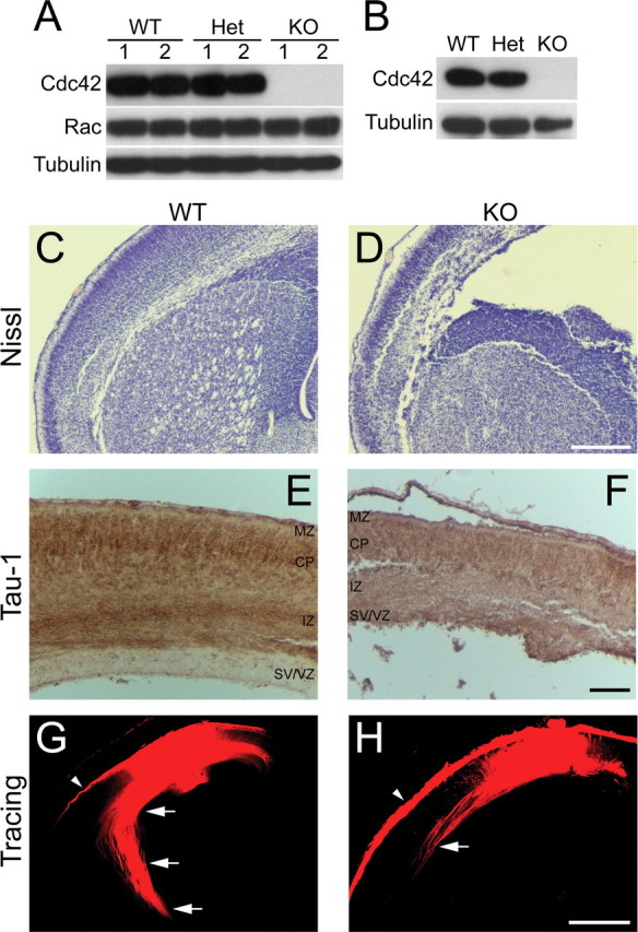
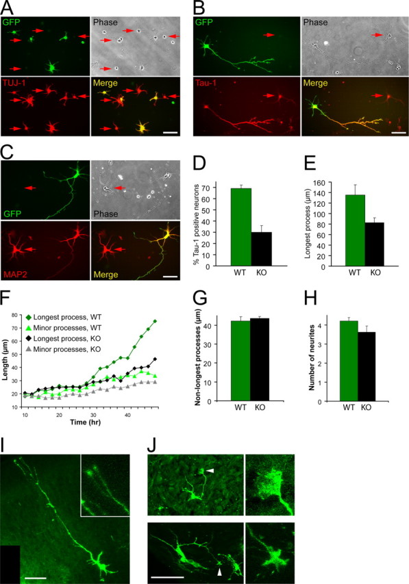
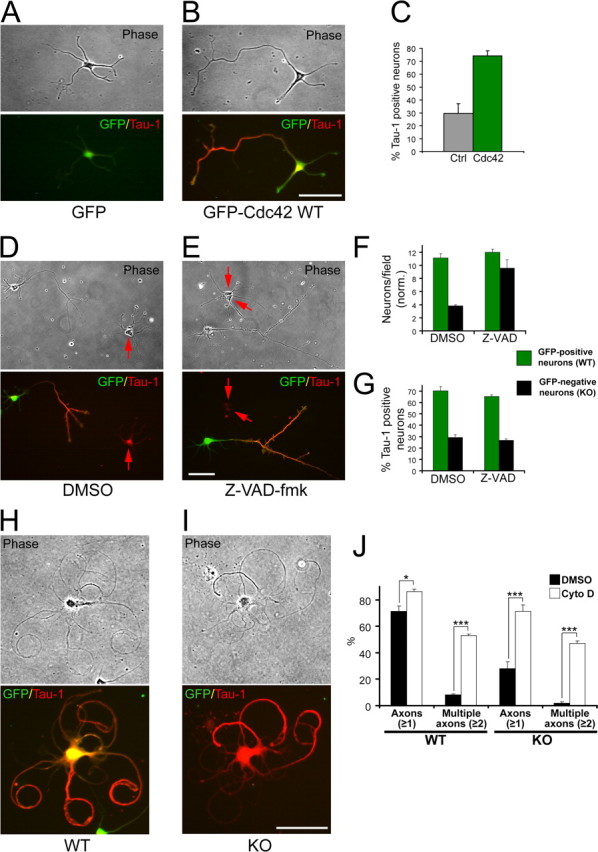
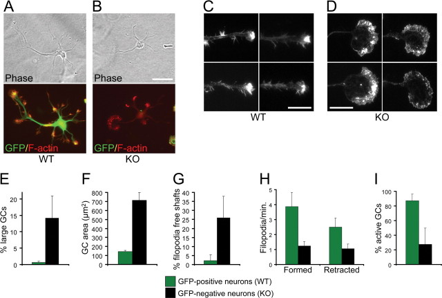
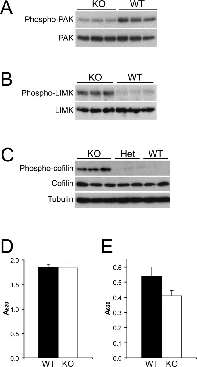
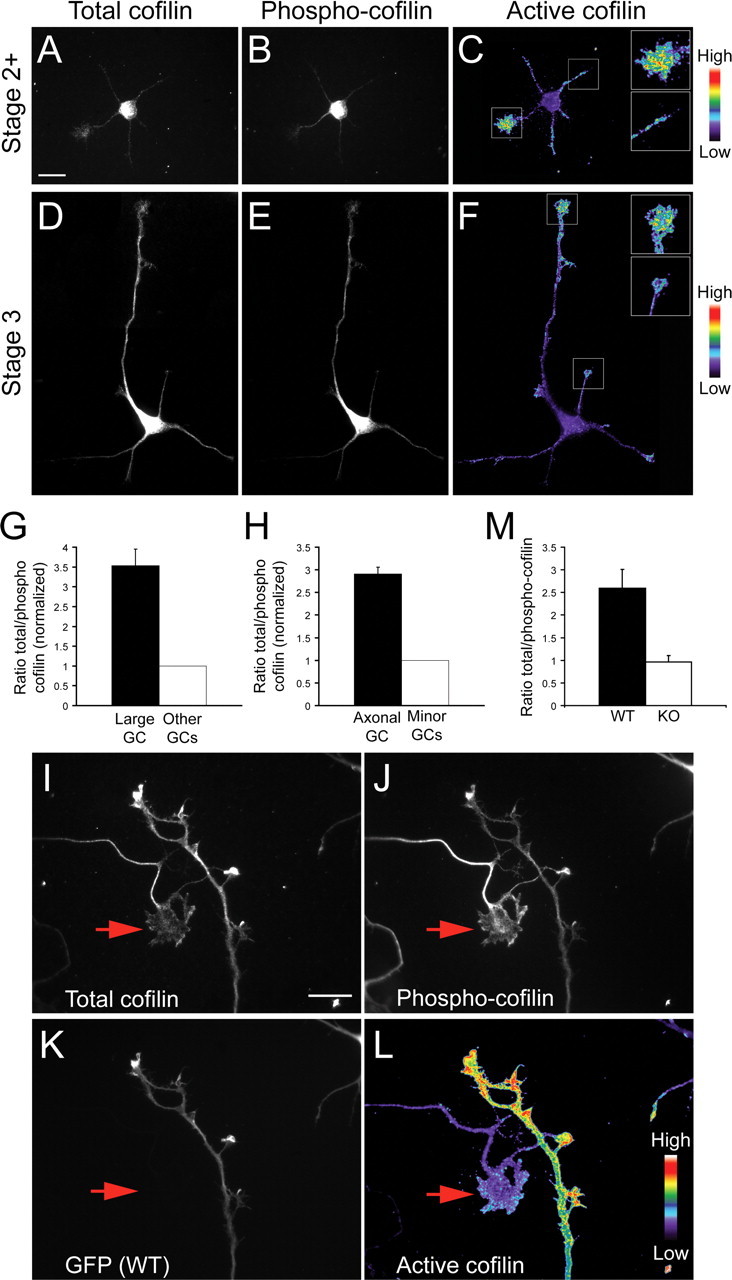
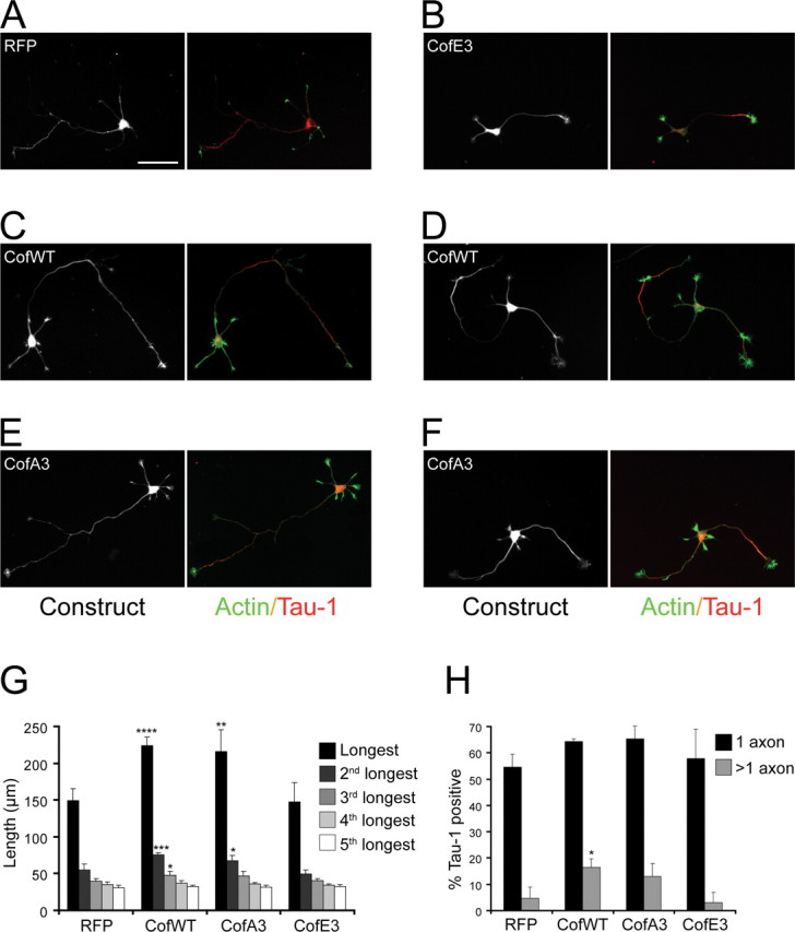
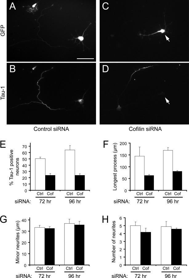
The establishment of polarity is an essential process in early neuronal development. Although a number of molecules controlling neuronal polarity have been identified, genetic evidence about their physiological roles in this process is mostly lacking. We analyzed the consequences of loss of Cdc42, a central regulator of polarity in multiple systems, on the polarization of mammalian neurons. Genetic ablation of Cdc42 in the brain led to multiple abnormalities, including striking defects in the formation of axonal tracts. Neurons from the Cdc42 null animals sprouted neurites but had a strongly suppressed ability to form axons both in vivo and in culture. This was accompanied by disrupted cytoskeletal organization, enlargement of the growth cones, and inhibition of filopodial dynamics. Axon formation in the knock-out neurons was rescued by manipulation of the actin cytoskeleton, indicating that the effects of Cdc42 ablation are exerted through modulation of actin dynamics. In addition, the knock-outs showed a specific increase in the phosphorylation (inactivation) of the Cdc42 effector cofilin. Furthermore, the active, nonphosphorylated form of cofilin was enriched in the axonal growth cones of wild-type, but not of mutant, neurons. Importantly, cofilin knockdown resulted in polarity defects quantitatively analogous to the ones seen after Cdc42 ablation. We conclude that Cdc42 is a key regulator of axon specification, and that cofilin is a physiological downstream effector of Cdc42 in this process.


Figures

Figure 1.
Figure 1.
Cdc42 depletion in vivo. A, B, Depletion of the Cdc42 protein in embryonic cortices and hippocampi. Mice double heterozygous for nestin-Cre and a floxed Cdc42 allele (Nes-Cre +/−, Cdc42 fl/wt) were crossed with homozygously floxed Cdc42 mice (Cdc42 fl/fl). The resulting embryos were dissected at E16.5, and protein extracts were prepared from the brain cortex (A) or from isolated hippocampi (B). Equal amounts of protein from each embryo were loaded on an SDS-PAGE and analyzed by Western blot with antibodies against Cdc42 and Rac1. A tubulin antibody was used as a control for equal loading. The Western blot in A shows the protein levels in two separate wild-type embryos (WT; Nes-Cre −/−, Cdc42 fl/wt or Nes-Cre −/−, Cdc42 fl/fl), two separate heterozygous embryos (Het; Nes-Cre +/−, Cdc42 fl/wt), and two separate Cdc42 knock-out embryos (KO; Nes-Cre +/−, Cdc42 fl/fl). The blots shown are representative of at least three independent experiments. C, D, Nissl staining of paraffin sections of E18.5 embryonic brains. Coronal sections at the level of the striatum of wild-type (C) and knock-out (D) brains are shown. Scale bar, 200 μm. E, F, Immunohistochemical staining of paraffin-embedded, E18.5 coronal sections with a Tau-1 antibody. Abundant Tau-1-positive axonal tracts are seen in the intermediate zone (IZ) in wild-type cortices (E), but very few are apparent in knock-out animals (F. Thinning and disorganization are also observed in other cortical layers, including the marginal zone (MZ), cortical plate (CP), and subventricular/ventricular zones (SV/VZ). Scale bar, 100 μm. G, H, DiI tracing of cortical axonal tracts. Fixed brains from wild-type (G) and knock-out (H E16.5 embryos were injected with DiI crystals. After diffusion of the dye, the labeled fibers in coronal sections at the level of the striatum were imaged by confocal microscopy. The axonal tracts were visualized inside the cortex (arrows); some surface labeling resulted from dye diffusion into the pia mater (arrowheads). The images shown are representative of at least five injected brains of each genotype; the axonal tracts were shorter and sparser in all knock-out animals in all regions of the cortex, compared with the wild-type counterparts. Scale bar, 500 μm.
Figure 2.
Figure 2.
Analysis of the phenotype of Cdc42 knock-out hippocampal neurons. Hippocampal neurons from Cdc42 knock-out E16.5 embryos were dissociated and mixed with wild-type hippocampal neurons from E16.5 embryos, ubiquitously expressing EGFP under a “CAG” promoter (Okabe et al., 1997; Ikawa et al., 1998), resulting in mixed cultures of GFP-positive wild-type neurons and GFP-negative Cdc42 knock-out neurons. A, Mixed cultures were fixed 10 h postplating and stained with an antibody against TUJ-1 to visualize all neuronal cells. B, Mixed cultures were fixed at 3 d postplating and stained with an antibody against a dephosphorylated epitope of Tau (Tau-1) to visualize axons. C, Mixed cultures were fixed at 3 d postplating and stained with an antibody against MAP2. The arrows in A–C indicate GFP-negative neurons, derived from Cdc42 knock-out hippocampi. All GFP-positive neurons are wild type. D, Quantification of the neurons having a Tau-1-positive process in mixed cultures. Green bars represent wild-type GFP-positive neurons, and black bars represent Cdc42 knock-out GFP-negative neurons. The data are from five separate cultures (number of cells quantified n ≥ 1200 per data point; p < 0.001). The values on this and all other graphs are averages plus SDs. E, Quantification of the length of the axon/longest neurite in mixed cultures (5 separate cultures; n ≥ 660 per data point; p < 0.001). F, Time course of process growth of wild-type and Cdc42 knock-out neurons imaged live. The graph shows the average length of the longest and minor (nonlongest) processes of eight wild-type and nine knock-out neurons. G, Length of the minor (nonlongest) processes of wild-type and Cdc42 knock-out neurons (3 separate cultures; n = 75 per data point; p = 0.41). H, Number of the neurites of wild-type and Cdc42 knock-out neurons (3 separate cultures; n = 75 per data point; p = 0.07). I, J, Brain sections of E18.5 wild-type (I and Cdc42 knock-out (J embryos were labeled with DiO using a ballistic delivery method to reveal individual neurons in the cortex. The inset in I shows a higher magnification of the wild-type growth cones. The arrowheads in J indicate enlarged growth cones in the Cdc42 knock-out neurons, which are shown at higher magnification on the right. Scale bars, 50 μm.
Figure 3.
Figure 3.
The polarization defects are rescued by Cdc42 re-expression and actin depolymerization but are not affected by inhibition of apoptosis. A, B, Cdc42 knock-out neurons were transfected with GFP-tagged wild-type Cdc42 (B) or with GFP (A) and stained for the axonal marker Tau-1. C, Quantification of the percentage of Cdc42 knock-out neurons having a Tau-1-positive process after transfection with control vector or with GFP-tagged wild-type Cdc42 (3 separate cultures; n ≥ 333; p < 0.001). D, E, Mixed wild-type (GFP-positive)/Cdc42 knock-out (GFP-negative) neuronal cultures were treated with DMSO (D) or with the general caspase inhibitor Z-VAD-fmk (E). The cultures were fixed 3 d later and stained for Tau-1. F, Quantification of the average number of neurons per field (normalized) in cultures treated with DMSO or with Z-VAD-fmk (≥60 fields counted per data point). G, Quantification of the neurons having a Tau-1-positive process in cultures treated with DMSO or with Z-VAD-fmk. The error bars are SDs (n ≥ 600 per data point). The data in F and G are representative of three separate cultures. H, I, Mixed cultures of wild-type GFP-positive neurons (H and Cdc42 knock-out GFP-negative neurons (I were treated with cytochalasin D 24 h postplating, fixed at 3 d, and stained for Tau-1. Scale bars, 50 μm. J, Quantification of the wild-type and knock-out neurons having processes positive for Tau-1 (Axons ≥1) or multiple Tau-1-positive axons (Multiple axons ≥2). The black bars represent DMSO-treated control cultures, and the white bars represent cytochalasin D-treated cultures (3 separate cultures; n ≥ 1250 per data point; *p < 0.05, ***p < 0.001).
Figure 4.
Figure 4.
Effects of Cdc42 ablation on the actin cytoskeleton. A, B, Mixed cultures of wild-type GFP-positive neurons (A) and Cdc42 knock-out GFP-negative neurons (B) were fixed at 3 d and stained with rhodamine-phalloidin to visualize actin filaments. Scale bar, 50 μm. C, D, Morphology of growth cones from wild-type neurons (C) and Cdc42 knock-out neurons (D) at 3 d in culture, visualized by rhodamine-phalloidin staining. Scale bars, 20 μm. E, Quantification of the neurons showing enlarged (>15 μm diameter) and smooth (filopodia-free) growth cones (5 separate cultures; n ≥ 660 per data point; p < 0.001). F, Quantification of the area of the enlarged Cdc42 knock-out growth cones compared with the area of wild-type growth cones (3 separate cultures; n ≥ 18; p < 0.001). G, Quantification of the neurons with smooth (filopodia-free) axonal/neurite shafts (5 separate cultures; n ≥ 660 per data point; p < 0.001). H, Quantification of the number of filopodia (thin protrusions longer than 3 μm) formed and retracted by wild-type and Cdc42 knock-out neurons per minute in time-lapse observations of live neurons (4 separate cultures; n ≥ 11 per data point; p < 0.01 for newly formed filopodia; p < 0.02 for retracted filopodia). I, Quantification of the percentage of growth cones showing rapid dynamics of filopodial/lamellipodial extension and retraction (active growth cones; 4 separate cultures; n ≥ 11 per data point; p < 0.01).
Figure 5.
Figure 5.
The level of cofilin phosphorylation in Cdc42 knock-out neurons is increased. A–C, Protein extracts from several separate E18.5 wild-type (WT), heterozygous (Het), and Cdc42 knock-out (KO) cortices were prepared and analyzed in parallel by Western blot. A, Cortical extracts were probed with an antibody specific for the phosphorylated Ser198/203 residues of PAK1 (top panel) and with a general anti-PAK antibody (bottom panel). B, Cortical extracts were probed with an antibody specific for the phosphorylated Thr508/505 residue of LIM kinase 1/2 (top panel) and with a general anti-LIM kinase (LIMK) antibody (bottom panel). C, Cortical extracts were probed with an antibody specific for the phosphorylated Ser3 residue of cofilin (top panel), with a general anti-cofilin antibody (middle panel), and with an anti-tubulin antibody as a loading control (bottom panel). D, Phosphatase activity in cortical extracts of wild-type (black bars) and Cdc42 knock-out embryos (white bars), measured with a generic phospho-Ser/Thr peptide substrate. The error bars are SDs from quadruplicate experiments (p > 0.5). E, Phosphatase activity in cortical extracts of wild-type (black bars) and Cdc42 knock-out (white bars) embryos, measured with a phospho-cofilin (Ser3) peptide substrate. The error bars are SDs from quadruplicate experiments (p < 0.02).
Figure 6.
Figure 6.
Active cofilin is enriched in axonal growth cones. Mouse hippocampal neurons at stage 2+ (A–C) or stage 3 (D–F) of development were fixed and stained with antibodies against total cofilin (A, D) and Ser3-phosphorylated cofilin (B, E). The ratio of the total cofilin to the phosphorylated cofilin, reflecting the relative amount of active cofilin, is depicted in C and F using a pseudocolor intensity scale. Insets show higher magnifications of the larger stage 2+ growth cones (C) or of the axonal growth cones at stage 3 (F, along with the next brightest growth cone. Scale bar, 20 μm. G, Quantification of the average cofilin/phospho-cofilin ratio in the larger growth cones of stage 2+ neurons (black bar) and in the remaining growth cones (open bar; 3 experiments; n ≥ 70; p < 0.001). H, Quantification of the average cofilin/phospho-cofilin ratio in the axonal growth cone of stage 3 neurons (black bar) and in the growth cones of minor neurites (open bar; 3 experiments; n ≥ 60; p < 0.001). I–L, Mixed cultures of wild-type hippocampal neurons expressing GFP (K and Cdc42 null neurons (GFP-negative) were stained for total cofilin (I and phospho-cofilin (J. The intensity ratio of the total cofilin/phospho-cofilin staining is shown in a pseudocolor scale in L. The arrow indicates the growth cone of the longest process of a Cdc42 knock-out (GFP-negative) neuron. Scale bar, 20 μm. M, Quantification of the total/phospho-cofilin ratio in the growth cones of wild-type and Cdc42 null growth cones (4 experiments; n ≥ 60; p < 0.001).
Figure 7.
Figure 7.
Wild-type and unphosphorylatable cofilin enhance axon growth. Mouse hippocampal neurons were infected with adenoviruses expressing RFP (A) or RFP fused to cofilinE3 (B) and cofilinA3 mutants (E, F or to cofilin wild-type (C, D). The cells were stained with phalloidin to reveal actin filaments and with a Tau-1 antibody to label the axons. Scale bar, 50 μm. The length of the individual processes (G) and the percentage of cells having Tau-1 positive axons (H were quantified (3–4 cultures; n ≥ 35; *p ≤ 0.05, **p ≤ 0.01, ***p ≤ 0.005, ****p ≤ 0.001).
Figure 8.
Figure 8.
Cofilin is necessary for axon formation. A–D, Mouse hippocampal neurons were infected with adenoviruses expressing human cofilin siRNA (control) (A, B) or mouse cofilin siRNA (C, D). A and C show the GFP signal (GFP was coexpressed as a reporter for the presence of siRNA in the neurons); B and D show Tau-1 immunostaining of the infected neurons. The arrow in C and D indicates a neuron expressing cofilin siRNA. Scale bar, 50 μm. E, Quantification of the control infected neurons (Ctrl; open bars) and mouse cofilin siRNA infected neurons (Cof; black bars) with a single Tau-1 positive process (axon) measured at 72 and 96 h postinfection. The error bars are SDs from triplicate experiments at 72 h (n > 60; p < 0.01) and from duplicate experiments at 96 h (n ≥ 34; p < 0.02). F–H, Quantification of the average length of the longest neurite (F, the length of the minor (nonlongest) neurites (G), and the total number of neurites (H in control infected neurons (open bars) and mouse cofilin siRNA infected neurons (black bars). The error bars are SDs from triplicate experiments at 72 h (n > 60) and from quadruplicate experiments at 96 h (n > 89). For the length of the longest neurite, p < 0.001 at both 72 and 96 h; for the remaining measurements, p > 0.05.

References

    1. Abe H, Ohshima S, Obinata T. A cofilin-like protein is involved in the regulation of actin assembly in developing skeletal muscle. J Biochem (Tokyo) 1989;106:696–702. - PubMed
    1. Abe T, Kato M, Miki H, Takenawa T, Endo T. Small GTPase Tc10 and its homologue RhoT induce N-WASP-mediated long process formation and neurite outgrowth. J Cell Sci. 2003;116:155–168. - PubMed
    1. Agnew BJ, Minamide LS, Bamburg JR. Reactivation of phosphorylated actin depolymerizing factor and identification of the regulatory site. J Biol Chem. 1995;270:17582–17587. - PubMed
    1. Andersen SS, Bi GQ. Axon formation: a molecular model for the generation of neuronal polarity. BioEssays. 2000;22:172–179. - PubMed
    1. Andrianantoandro E, Pollard TD. Mechanism of actin filament turnover by severing and nucleation at different concentrations of ADF/cofilin. Mol Cell. 2006;24:13–23. - PubMed

Publication types

MeSH terms

Substances

LinkOut - more resources