. 2007 Nov 28;27(48):13303-10.
doi: 10.1523/JNEUROSCI.2349-07.2007.

Is the prefrontal cortex necessary for establishing cognitive sets?

Affiliations

Is the prefrontal cortex necessary for establishing cognitive sets?

James B Rowe et al. J Neurosci. .

Abstract

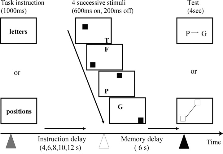
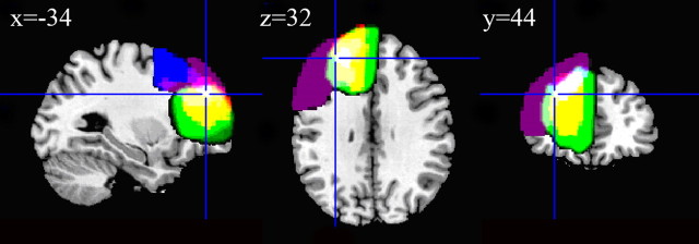
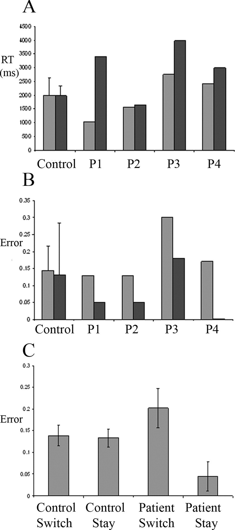
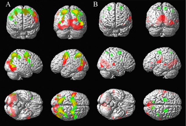
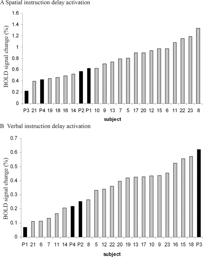
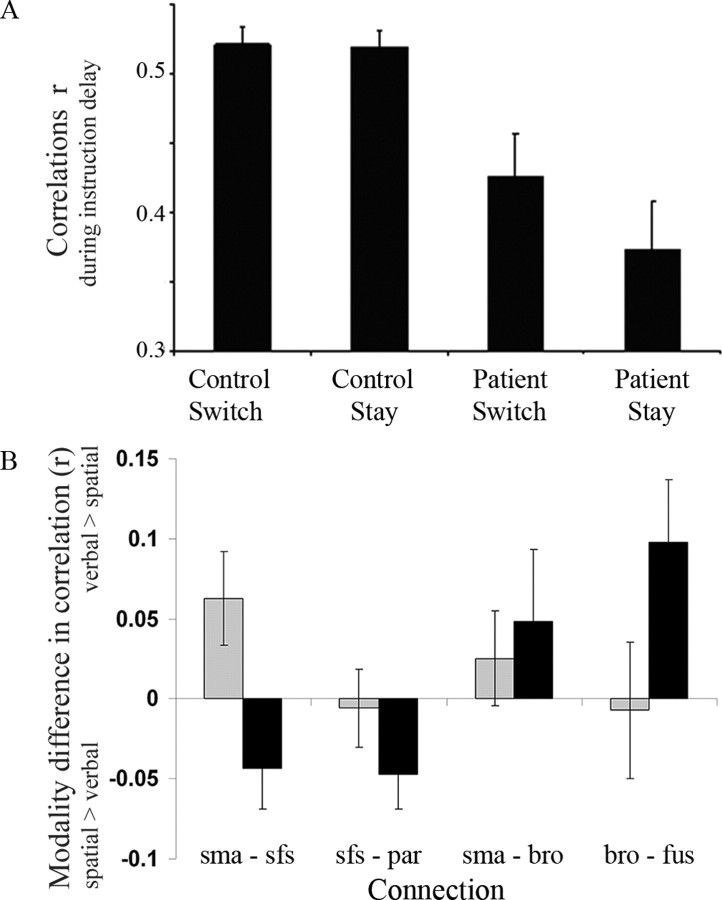
There is evidence from neuroimaging that the prefrontal cortex may be involved in establishing task set activity in advance of presentation of the task itself. To find out whether it plays an essential role, we examined patients with unilateral lesions of the rostral prefrontal cortex. They were first instructed as to whether to perform a spatial or a verbal working memory task and then given spatial and verbal items after a delay of 4-12 s. The patients showed an increase in switch costs, making more errors by repeating what they had done on the previous trial. They were able to establish regional task set activity during the instruction delay, as evidenced by sustained changes in the blood oxygenation level-dependent signal in caudal frontal regions. However, in contrast to healthy controls, they were less able to maintain functional connectivity among the surviving task-related brain regions, as evidenced by reduced correlations between them during instruction delays. The results suggest that the left rostral prefrontal cortex is indeed required for establishing a cognitive set but that the essential function is to support the functional connectivity among the task-related regions.


Figures

Figure 1.
Figure 1.
The task gave subjects advanced warning of the modality of a forthcoming verbal (letter-based) or spatial (position-based) working memory task allowing the subjects to establish the relevant cognitive set in advance of the actual stimuli. This “instruction delay” varied between 4 and 12 s. The stimuli each included a letter and square of specific location, but only one modality needed to be attended to and remembered. The order of letters or locations was remembered for 6 s and tested by a single probe cue that asked about the order of two of the four remembered stimuli (e.g., in these examples, was the letter P followed immediately by the letter G, or was the bottom left square followed immediately by the top square?) (yes to both examples).
Figure 2.
Figure 2.
The four patients' (P1–P4) lesions are shown overlaid on the canonical brain in MNI space. The four patients are separately color coded: P1, purple, overlapping as pink, white); P2, blue, overlapping as light pink and light purple, white); P3, green, overlapping as yellow, white); P4, red, overlapping as yellow, white). Complete overlap of all lesions is indicated by white. The cross-hairs are placed at the peak of prefrontal activation associated with set-related activity in previous studies (Sakai et al., 2006).
Figure 3.
Figure 3.
A, Reaction times (RT). B, Error rates for the control group and each patient (P1–P4). In A and B, spatial tasks are indicated by light gray bars, and verbal tasks are indicated by dark gray bars. C, Error rates are shown separately for trials in which the modality had just switched (switch) or was the same as the previous trial (stay), for control and patients groups, respectively, averaging across modality. Control data are shown as group mean ± SE.
Figure 4.
Figure 4.
SPM{t} maps of activations during the instruction delay, before subjects have seen the stimuli that are to be remembered. A, Thresholded at p < 0.05 (FWE) for verbal (red) and spatial (green) instruction delays against baseline. B, Thresholded at p < 0.001 (uncorrected) for the difference between modalities. Activations that were greater for verbal than spatial instruction delays are shown in red, whereas activations that were greater for spatial than verbal instruction delays are shown in green. See Results for corrected significance levels within ROIs. SPM{t}s are shown superimposed on a canonical T1-weighted image in MNI space.
Figure 5.
Figure 5.
The set activity in area 8 at −24, −8, 56 during spatial instruction delay (top) and in area 44 at −54, 04, 22 during verbal instruction delay (bottom) is plotted separately for each subject. The four patients (subjects P1–P4) are marked in black, and control subjects (subjects 5–23) are marked in gray. The instruction delay activity effect size is in arbitrary units indicating the BOLD signal change, derived from the contrast images of verbal instruction delay and spatial instruction delay, respectively.
Figure 6.
Figure 6.
A, The correlations (r) among the pre-SMA, area 44, area 8, and parietal and fusiform cortex were lower in patients, more so on stay trials (bars show group means averaged across all connections and both modalities ± SE). B, The modality difference in correlation r, between spatial and verbal instruction delays, varied between the four anatomical connections and depended on the switch–stay factor but not the group (see Results for ANOVA results). A positive value indicates higher correlations during verbal instruction periods, whereas a negative value indicates higher correlations during spatial instruction periods. Black bars represent stay trials, and gray bars represent switch trials. sma, Pre-SMA; sfs, superior frontal sulcus; par, parietal cortex; bro, area 44 of Broca's area; fus, fusiform gyrus.

Comment in

References

    1. Aron AR, Monsell S, Sahakian BJ, Robbins TW. A componential analysis of task-switching deficits associated with lesions of left and right frontal cortex. Brain. 2004;127:1561–1573. - PubMed
    1. Blair J, Spreen O. Predicting premorbid IQ: a revision of the National Adult Reading Test. Clin Neuropsychologist. 1989;3:129–136.
    1. Bunge SA, Kahn I, Wallis JD, Miller EK, Wagner AD. Neural circuits subserving the retrieval and maintenance of abstract rules. J Neurophysiol. 2003;90:3419–3428. - PubMed
    1. Burgess PW, Veitch E, de Lacy Costello A, Shallice T. The cognitive and neuroanatomical correlates of multitasking. Neuropsychologia. 2000;38:848–863. - PubMed
    1. Butters N, Pandya D, Sanders K, Dye P. Behavioral deficits in monkeys after selective lesions within the middle third of sulcus principalis. J Comp Physiol Psychol. 1971;76:8–14. - PubMed

Publication types

MeSH terms