. 2008 Feb;82(3):1622-5.
doi: 10.1128/JVI.02097-07. Epub 2007 Nov 28.

Murine endogenous retrovirus MuERV-L is the progenitor of the "orphan" epsilon viruslike particles of the early mouse embryo

Affiliations

Murine endogenous retrovirus MuERV-L is the progenitor of the "orphan" epsilon viruslike particles of the early mouse embryo

David Ribet et al. J Virol. 2008 Feb.

Abstract

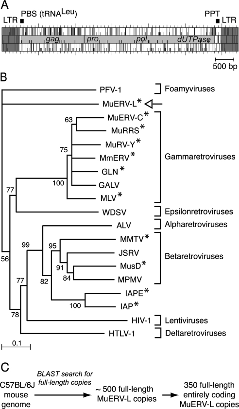
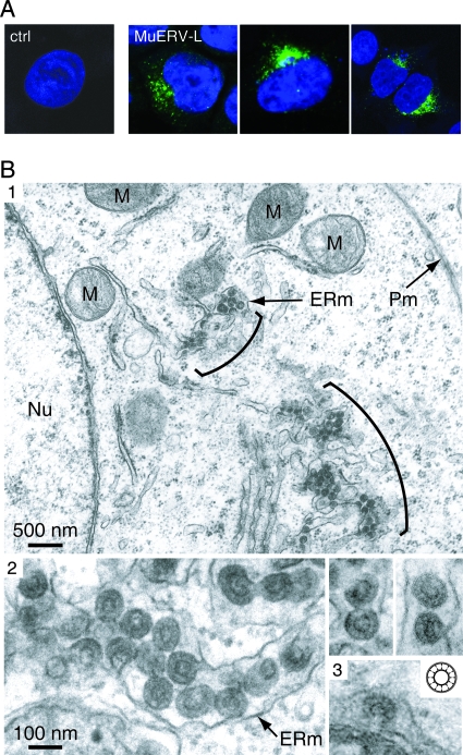
Viruslike particles which displayed a peculiar wheellike appearance that distinguished them from A-, B- or C-type particles had previously been described in the early mouse embryo. The maximum expression of these so-called epsilon particles was observed in two-cell-stage embryos, followed by their rapid decline at later stages of development and no particles detected at the zygote one-cell stage. Here, we show that these particles are in fact produced by a newly discovered murine endogenous retrovirus (ERV) belonging to the widespread family of mammalian ERV-L elements and named MuERV-L. Using antibodies that we raised against the Gag protein of these elements, Western blot analysis and in toto immunofluorescence studies of the embryos at various stages disclosed the same developmental expression profile as that observed for epsilon particles. Using expression vectors for cloned, full-length, entirely coding MuERV-L copies and cell transfection, direct identification of the epsilon particles was finally achieved by high-resolution electron microscopy.


Figures

FIG. 1.
FIG. 1.
Structure and phylogeny of MuERV-L elements. (A) Genomic organization of an MuERV-L provirus, with the LTRs (in dark gray) flanking two ORFs homologous to the retroviral gag and pol genes (in light gray). The pol gene further discloses domains homologous to retroviral pro and dUTPase genes. A primer binding site (PBS) complementary to tRNALeu and a polypurine track (PPT) can be identified. (B) Phylogeny of retroviruses, based on their reverse transcriptase domain. The tree was constructed by the neighbor-joining method using the seven blocks of conserved residues found in the reverse transcriptase domain of all retroelements and was rooted with non-LTR retrotransposons. All sequences are readily accessible from GenBank and previous reports (e.g., reference 15). Percent bootstrap values obtained from 1,000 replicates are indicated. The retroviruses “endogenized” in the mouse genome are marked (*). The seven retroviral genera are indicated on the right. PFV, primate foamy virus; MuRRS, murine retrovirus-related DNA sequence; MuRV-Y, murine retrovirus Y associated; MmERV, Mus musculus ERV; GLN, murine retrovirus using tRNAGln; GALV, gibbon ape leukemia virus; MLV, murine leukemia virus; WDSV, walleye dermal sarcoma virus; ALV, avian leukosis virus; MMTV, mouse mammary tumor virus; JSRV, jaagsiekte sheep retrovirus; MusD, Mus musculus type D retrovirus; MPMV, Mason-Pfizer monkey virus; IAPE, IAP with an envelope gene; HIV-1, human immunodeficiency virus type 1; HTLV-1, human T-cell leukemia virus type 1. (C) Search for full-length, entirely coding MuERV-L copies in the C57BL/6J genome. Blast search was carried out with the NCBI m36 mouse assembly (April 2006 release) and yielded 489 full-length MuERV-L copies among which 350 contain the two complete gag and pol ORFs.
FIG. 2.
FIG. 2.
Expression of the MuERV-L Gag protein in the early mouse embryo. (A) Western blot analysis with a rabbit anti-Gag MuERV-L antiserum of germinal vesicles (GV); two-cell stage (2c), eight-cell stage (8c), and blastocyst stage (Bl) mouse embryos (left panel); or human 293T cells transfected with the MuERV-L expression vector or a control (ctrl) plasmid (right panel): the lanes in the left panel correspond to an amount of 60 GV or mouse embryos, collected from mated Swiss (OF1) mice and lysed in Laemmli buffer for sodium dodecyl sulfate-polyacrylamide gel electrophoresis and Western blotting. (B) Immunofluorescence confocal analysis of germinal vesicles (GV) and two-cell stage (2c) and eight-cell stage (8c) mouse embryos. The samples were fixed in 4% formaldehyde, permeabilized, and stained with the anti-Gag MuERV-L antiserum and a fluorescein isothiocyanate-conjugated antirabbit immunoglobulin G secondary antibody. Preincubation of the antiserum (30 min at 4°C) with an excess of recombinant MuERV-L Gag protein before staining of the two-cell stage embryos was carried out as a control (2c + Gag).
FIG. 3.
FIG. 3.
Localization and morphology of MuERV-L-associated viruslike particles. (A) Confocal images of human cells transfected with the MuERV-L vector or a control plasmid (ctrl). Forty-eight hours posttransfection, cells were fixed, permeabilized, and stained with the anti-Gag MuERV-L antiserum and an Alexa fluor 488-conjugated antirabbit immunoglobulin G secondary antibody (in green). Nuclei were stained with TO-PRO-3 iodide (in blue). (B) Electron microscopy of mouse epsilon particles. (1) Representative low-magnification image of 293T cells transfected with MuERV-L disclosing particles accumulated in the cisternae of the ER (brackets). No particles can be observed at the plasma membrane (Pm). ERm, ER membrane; Nu, nucleus; M; mitochondria. (2) High-magnification images of 293T cells transfected with MuERV-L disclosing particles within or budding into the ER. (3) High-magnification image of a naturally occurring epsilon particle budding into the ER of a mouse embryo, and schematic structure of an epsilon particle (inset, adapted with permission from reference 13).

References

    1. Bénit, L., N. de Parseval, J.-F. Casella, I. Callebaut, A. Cordonnier, and T. Heidmann. 1997. Cloning of a new murine endogenous retrovirus, MuERV-L, with strong similarity to the human HERV-L element and with a gag coding sequence closely related to the Fv1 restriction gene. J. Virol. 715652-5657. - PMC - PubMed
    1. Bénit, L., J. B. Lallemand, J. F. Casella, H. Philippe, and T. Heidmann. 1999. ERV-L elements: a family of endogenous retrovirus-like elements active throughout the evolution of mammals. J. Virol. 733301-3308. - PMC - PubMed
    1. Bernhard, W. 1958. Electron microscopy of tumor cells and tumor viruses; a review. Cancer Res. 18491-509. - PubMed
    1. Boeke, J. D., and J. P. Stoye. 1997. Retrotransposons, endogenous retroviruses, and the evolution of retroelements, p. 343-435. In J. M. Coffin, S. H. Hughes, and H. E. Varmus (ed.), Retroviruses. Cold Spring Harbor Laboratory Press, Cold Spring Harbor, NY. - PubMed
    1. Cordonnier, A., J.-F. Casella, and T. Heidmann. 1995. Isolation of novel human endogenous retrovirus-like elements with foamy virus-related pol sequence. J. Virol. 695890-5897. - PMC - PubMed

Publication types

MeSH terms

LinkOut - more resources