. 2007 Dec 15;93(12):4453-61.
doi: 10.1529/biophysj.106.101386.

Fibroblast adaptation and stiffness matching to soft elastic substrates

Affiliations

Fibroblast adaptation and stiffness matching to soft elastic substrates

Jérôme Solon et al. Biophys J. .

Abstract

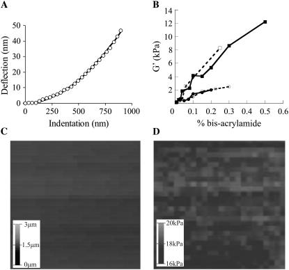
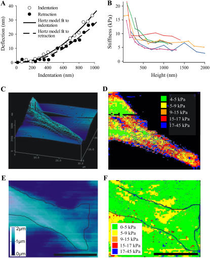
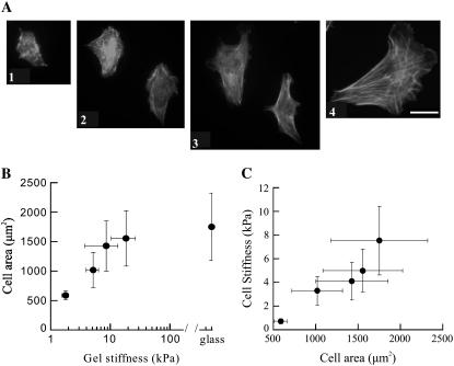
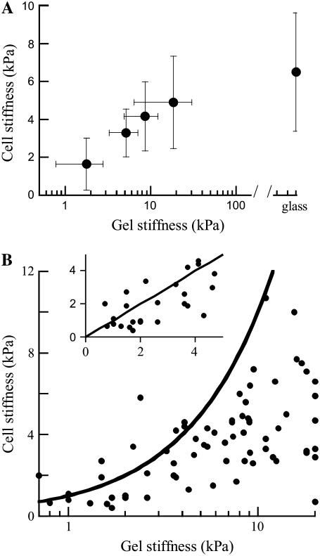
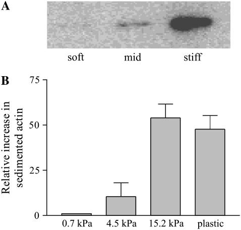
Many cell types alter their morphology and gene expression profile when grown on chemically equivalent surfaces with different rigidities. One expectation of this change in morphology and composition is that the cell's internal stiffness, governed by cytoskeletal assembly and production of internal stresses, will change as a function of substrate stiffness. Atomic force microscopy was used to measure the stiffness of fibroblasts grown on fibronectin-coated polyacrylamide gels of shear moduli varying between 500 and 40,000 Pa. Indentation measurements show that the cells' elastic moduli were equal to, or slightly lower than, those of their substrates for a range of soft gels and reached a saturating value at a substrate rigidity of 20 kPa. The amount of cross-linked F-actin sedimenting at low centrifugal force also increased with substrate stiffness. Together with enhanced actin polymerization and cross-linking, active contraction of the cytoskeleton can also modulate stiffness by exploiting the nonlinear elasticity of semiflexible biopolymer networks. These results suggest that within a range of stiffness spanning that of soft tissues, fibroblasts tune their internal stiffness to match that of their substrate, and modulation of cellular stiffness by the rigidity of the environment may be a mechanism used to direct cell migration and wound repair.


Figures

FIGURE 1
FIGURE 1
Polyacrylamide gel characterization by AFM. (A) Representative curve of cantilever deflection as a function of tip indentation into a 5-kPa PA gel (circles) and the fit to the data with the Hertz model (bold line). (B) PA gel stiffness as a function of bis-acrylamide cross-linker concentration (open squares/dashed line, AFM 7.5% acrylamide; solid squares/solid line, macroscopic rheology 7.5% acrylamide; open circles/dashed line, AFM 5% acrylamide (8); solid circles/solid line, macroscopic rheology 5% acrylamide). (C) Topographical map of the surface of a 5-kPa PA gel. Scale, 30 μm × 30 μm. (D) Stiffness map of the surface of a 5-kPa PA gel. Scale, 30 μm × 30 μm.
FIGURE 2
FIGURE 2
Fibroblast characterization by AFM. (A) Representative curve of cantilever deflection as a function of tip indentation into (open circles) and retraction from (solid circles) a fibroblast plated on a fibronectin-laminated 5-kPa PA gel and the fit to the data with the Hertz model (solid line, indentation; dashed line, retraction). (B) Variation in measured cell stiffness as a function of cell height from the substrate (fibronectin-coated glass) for several individual cells. Each curve represents a single cell. (C) Topographical map of the edge of a fibroblast adhering to a glass substrate. Scale, 30 μm × 30 μm. (D) Stiffness map of the edge of a fibroblast adhering to a glass substrate coated with fibronectin. Scale, 30 μm × 30 μm. (E) Topographical map of the edge of a fibroblast adhering to a fibronectin-laminated 5-kPa PA gel. Bold line shows the outline of the cell. Scale bar = 10 μm. (F) Stiffness map of the edge of a fibroblast adhering to a fibronectin-laminated 5-kPa PA gel. Scale bar = 10 μm.
FIGURE 3
FIGURE 3
Microscopic analysis of fibroblasts on gels. (A) Rhodamine-phalloidin staining of the F-actin of fixed fibroblasts on a 1-kPa gel (1), 5-kPa gel (2), 10-kPa gel (3), and glass (4). Bar, 40 μm. (B) Projected cell area as a function of gel stiffness. Each point on the graph is a mean ± SD of 12–40 different cells. (C) Cell stiffness as a function of cell area. Each point on the graph is a mean ± SD of 12–40 different cells.
FIGURE 4
FIGURE 4
Effect of substrate stiffness on cell stiffness. (A) Cell stiffness as a function of the stiffness of the adjacent gel. Each point is a mean ± SD of 12–40 different cells. (B) Individual measurements of cell stiffness as a function of adjacent gel stiffness. Each point shown is the mean stiffness of a single cell plotted against that of the neighboring gel. Bold line is the line of identity showing the gel stiffness. The inset is an enlargement of the range of gel stiffness up to 5 kPa on a linear scale.
FIGURE 5
FIGURE 5
Effect of substrate stiffness of F-actin organization. (A) Western blot for actin from pellet formed by low-speed (15,000 × g) centrifugation of fibroblast lysates. Cells were lysed 24 h after plating on fibronectin-laminated PA gels with 0.7 kPa, 4.5 kPa, and 15.2 kPa elastic moduli. (B) Densitometric quantification of Western blots for actin sedimenting at low speed from fibroblasts plated on PA gels of 0.7 kPa, 4.5 kPa, 15.2 kPa, or tissue culture plastic. All values normalized to total protein in cell lysate. Error bars are representative standard deviations from three repeats.

References

    1. Levental, I., P. C. Georges, and P. A. Janmey. 2007. Soft biological materials and their impact on cell function. Soft Matter. 1:299–306. - PubMed
    1. Engler, A. J., S. Sen, H. L. Sweeney, and D. E. Discher. 2006. Matrix elasticity directs stem cell lineage specification. Cell. 126:677–689. - PubMed
    1. Pelham, R. J., Jr., and Y. Wang. 1997. Cell locomotion and focal adhesions are regulated by substrate flexibility. Proc. Natl. Acad. Sci. USA. 94:13661–13665. - PMC - PubMed
    1. Discher, D. E., P. Janmey, and Y. L. Wang. 2005. Tissue cells feel and respond to the stiffness of their substrate. Science. 310:1139–1143. - PubMed
    1. Georges, P. C., and P. A. Janmey. 2005. Cell type-specific response to growth on soft materials. J. Appl. Physiol. 98:1547–1553. - PubMed

LinkOut - more resources