. 2008 Feb;19(2):587-94.
doi: 10.1091/mbc.e07-10-1051. Epub 2007 Nov 28.

KNL1 and the CENP-H/I/K complex coordinately direct kinetochore assembly in vertebrates

Affiliations

KNL1 and the CENP-H/I/K complex coordinately direct kinetochore assembly in vertebrates

Iain M Cheeseman et al. Mol Biol Cell. 2008 Feb.

Abstract

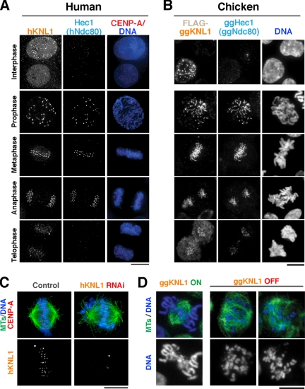
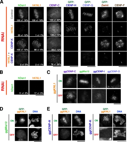
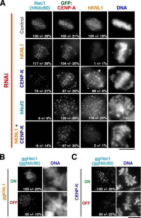
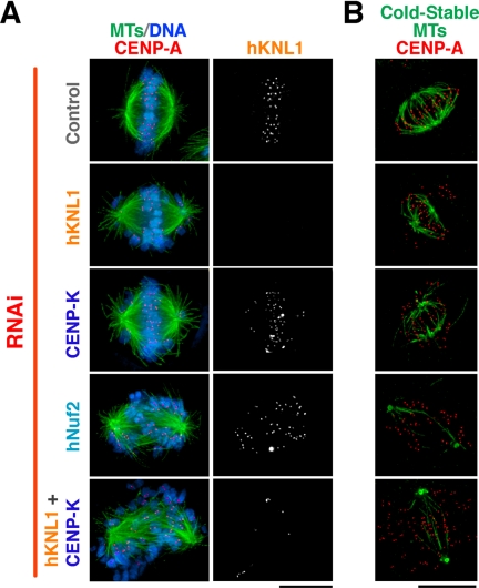
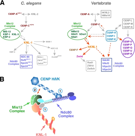
Chromosome segregation during mitosis requires the assembly of a large proteinaceous structure termed the kinetochore. In Caenorhabditis elegans, KNL-1 is required to target multiple outer kinetochore proteins. Here, we demonstrate that the vertebrate KNL1 counterpart is essential for chromosome segregation and is required to localize a subset of outer kinetochore proteins. However, unlike in C. elegans, depletion of vertebrate KNL1 does not abolish kinetochore localization of the microtubule-binding Ndc80 complex. Instead, we show that KNL1 and CENP-K, a subunit of a constitutively centromere-associated complex that is missing from C. elegans, coordinately direct Ndc80 complex localization. Simultaneously reducing both hKNL1 and CENP-K function abolishes all aspects of kinetochore assembly downstream of centromeric chromatin and causes catastrophic chromosome segregation defects. These findings explain discrepancies in kinetochore assembly pathways between different organisms and reveal a surprising plasticity in the assembly mechanism of an essential cell division organelle.


Figures

Figure 1.
Figure 1.
Vertebrate KNL1 localizes to kinetochores during mitosis and is required for chromosome segregation. hKNL1 and ggKNL1 localize to kinetochores during mitosis. (A) Images showing hKNL1, Hec1 (hNdc80), stably expressed GFP-CENP-A (red), and DNA (blue) throughout the cell cycle in human cells. (B) Images showing FLAG-ggKNL1, ggHEC1 (ggNdc80), and DNA throughout the cell cycle. (C) hKNL1 depletion results in chromosome misalignment. Immunofluorescence images of control siRNA-transfected or hKNL1 RNAi-transfected cells stained for microtubules (green), GFP-CENP-A (red), DNA (blue), and hKNL1. (D) ggKNL1 depletion results in chromosome misalignment. Immunofluorescence images of conditional ggKNL1 DT40 cell lines either in the absence of tetracycline (ON) or 36 h after the addition of tetracycline (OFF) showing microtubules and DNA. See Supplementary Figure 1 for details on the cell line and extent of depletion. Scale bars, 10 μm.
Figure 2.
Figure 2.
Localization of kinetochore proteins after KNL1/CENP-H/IK inhibitions. (A) Immunofluorescence images acquired using antibodies to hDsn1, hKNL1, CENP-C, and CENP-F, or using anti-GFP antibody in cells stably expressing GFP-CENP-H, GFP-CENP-Q, and GFP-Zwint. Control cells, hKNL1-depleted cells, CENP-K–depleted cells and cells doubly depleted for CENP-K and hKNL1 are shown. (B) hDsn1-depleted cells costained for hDsn1 and hKNL1 as in A. In A and B, when partial defects in localization were evident, kinetochore fluorescence intensity was measured relative to control cells. The number in bottom right represents the average kinetochore fluorescence intensity ± SD expressed as a percentage relative to control cells. Intensities were measured for between four and six cells, with 30–80 kinetochores per cell for each condition. (C) Immunofluorescence images of conditional ggKNL1 DT40 cell lines either in the absence of tetracycline (ON) or 36 h after the addition of tetracycline (OFF), showing ggCENP-C, ggMis12, ggCENP-I, or ggCENP-O localization detected using specific antibodies. (D) Immunofluorescence images of conditional ggMis12 DT40 cell lines either in the absence of tetracycline (ON) or 36 h after the addition of tetracycline (OFF), showing localization of a stably transfected GFP-ggKNL1 fusion protein. (E). Immunofluorescence images of conditional ggCENP-H and ggCENP-K DT40 cell lines either in the absence of tetracycline (ON) or 36 h after the addition of tetracycline (OFF), showing localization of a stably transfected GFP-ggKNL1 fusion protein. Scale bars, 10 μm.
Figure 3.
Figure 3.
KNL1 and CENP-K act coordinately to recruit the Ndc80 complex to kinetochores. (A) Immunofluorescence images acquired using antibodies against Hec1 (hNdc80), hKNL1, and an anti-GFP antibody in cells stably expressing GFP-CENP-A. Control cells, hKNL1-depleted cells, CENP-K–depleted cells, hNuf2-depleted cells, and cells doubly depleted for CENP-K and hKNL1 are shown. Kinetochore fluorescence intensities were quantified in all channels and the mean ± SD, expressed as a percentage relative to control cells, is indicated in the bottom right. The fluorescence intensity of 30–80 kinetochores per cell was measured for between 12 and 23 cells per condition. (B and C) Immunofluorescence images showing ggHec1 (ggNdc80) localization in conditional ggKNL1 (B) or ggCENP-K (C) DT40 cell lines either in the absence of tetracycline (ON) or 36 h after the addition of tetracycline (OFF). Numbers in panels indicate mean ± SD in kinetochore fluorescence intensity of ggHec1 (ggNdc80) relative to the control ON cells. Scale bars, 10 μm.
Figure 4.
Figure 4.
Simultaneous depletion of hKNL1 and CENP-K results in catastrophic defects in chromosome segregation. (A) Microtubules (green), DNA (blue), CENP-A (red), and hKNL1 in control cells, hKNL1-depleted cells, CENP-K–depleted cells, hNuf2-depleted cells, and cells doubly depleted for CENP-K and hKNL1. (B) Microtubules (green) and CENP-A (red) staining after the indicated cells were incubated at 0°C for 10 min to selectively visualize cold stable kinetochore microtubule fibers. Scale bars, 10 μm.
Figure 5.
Figure 5.
Models for kinetochore assembly and synergistic contribution of KNL-1 and CENP-H/I/K to Ndc80 complex localization. (A) Diagram of kinetochore assembly hierarchies in C. elegans embryos and vertebrate cells. Red arrows indicate dependencies defined in this study. These schematics are not comprehensive and are focused primarily on stably associated proteins/complexes that do exhibit rapid turnover at kinetochores. (B) Schematic of potential mechanisms explaining the synergistic defect in Ndc80 complex localization in hKNL1/CENP-K double-depleted cells. The “two hand” model (1) is based on distinct sites for association of KNL1 and CENP-H/I/K on the four-subunit Ndc80 complex. Loss of a single association site would weaken, but not abolish, localization. Biochemical and two-hybrid studies provide some support for this idea. Partial localization interdependence between CENP-H/I/K and the Mis12 complex (2), which is known to affect Ndc80 complex localization in both C. elegans and vertebrates, may also indirectly contribute to the observed synergistic role of CENP-K and hKNL1 in Ndc80 complex localization. These two proposals are not mutually exclusive, and both are likely to contribute to some degree in vertebrates to ensure proper Ndc80 complex localization and function.

Similar articles

Cited by

References

    1. Cheeseman I. M., Chappie J. S., Wilson-Kubalek E. M., Desai A. The conserved KMN network constitutes the core microtubule-binding site of the kinetochore. Cell. 2006;127:983–997. - PubMed
    1. Cheeseman I. M., Niessen S., Anderson S., Hyndman F., Yates J. R., III, Oegema K., Desai A. A conserved protein network controls assembly of the outer kinetochore and its ability to sustain tension. Genes Dev. 2004;18:2255–2268. - PMC - PubMed
    1. De Wulf P., McAinsh A. D., Sorger P. K. Hierarchical assembly of the budding yeast kinetochore from multiple subcomplexes. Genes Dev. 2003;17:2902–2921. - PMC - PubMed
    1. DeLuca J. G., Gall W. E., Ciferri C., Cimini D., Musacchio A., Salmon E. D. Kinetochore microtubule dynamics and attachment stability are regulated by Hec1. Cell. 2006;127:969–982. - PubMed
    1. DeLuca J. G., Moree B., Hickey J. M., Kilmartin J. V., Salmon E. D. hNuf2 inhibition blocks stable kinetochore-microtubule attachment and induces mitotic cell death in HeLa cells. J. Cell Biol. 2002;159:549–555. - PMC - PubMed

Publication types

MeSH terms

LinkOut - more resources