. 2008 Feb;19(2):553-62.
doi: 10.1091/mbc.e07-09-0890. Epub 2007 Nov 28.

Myosin transducer mutations differentially affect motor function, myofibril structure, and the performance of skeletal and cardiac muscles

Affiliations

Myosin transducer mutations differentially affect motor function, myofibril structure, and the performance of skeletal and cardiac muscles

Anthony Cammarato et al. Mol Biol Cell. 2008 Feb.

Abstract

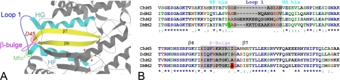
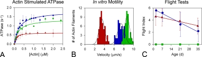
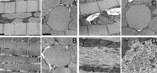
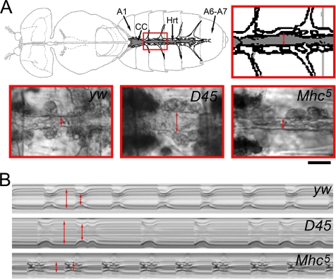
Striated muscle myosin is a multidomain ATP-dependent molecular motor. Alterations to various domains affect the chemomechanical properties of the motor, and they are associated with skeletal and cardiac myopathies. The myosin transducer domain is located near the nucleotide-binding site. Here, we helped define the role of the transducer by using an integrative approach to study how Drosophila melanogaster transducer mutations D45 and Mhc(5) affect myosin function and skeletal and cardiac muscle structure and performance. We found D45 (A261T) myosin has depressed ATPase activity and in vitro actin motility, whereas Mhc(5) (G200D) myosin has these properties enhanced. Depressed D45 myosin activity protects against age-associated dysfunction in metabolically demanding skeletal muscles. In contrast, enhanced Mhc(5) myosin function allows normal skeletal myofibril assembly, but it induces degradation of the myofibrillar apparatus, probably as a result of contractile disinhibition. Analysis of beating hearts demonstrates depressed motor function evokes a dilatory response, similar to that seen with vertebrate dilated cardiomyopathy myosin mutations, and it disrupts contractile rhythmicity. Enhanced myosin performance generates a phenotype apparently analogous to that of human restrictive cardiomyopathy, possibly indicating myosin-based origins for the disease. The D45 and Mhc(5) mutations illustrate the transducer's role in influencing the chemomechanical properties of myosin and produce unique pathologies in distinct muscles. Our data suggest Drosophila is a valuable system for identifying and modeling mutations analogous to those associated with specific human muscle disorders.


Figures

Figure 1.
Figure 1.
Locations of myosin transducer mutations and the alignment and sequence comparison of transducer elements from various myosin isoforms. (A) Ribbon diagram (prepared using the KiNG 1.39 [Kinemage, Next Generation, Durham, NC] interactive system for three-dimensional vector graphics) of a portion of the chicken myosin V (Protein Data Bank code 1W7J) motor domain near the nucleotide (shown as a ball and stick model) binding pocket, with specific components of the transducer region labeled. The locations of D. melanogaster D45 and Mhc5 mutations are shown relative to specific transducer elements. D45 is at the junction between the β-bulge and the seventh strand of the central β-sheet, whereas Mhc5 lies close to the junction between the HF helix and hypervariable loop 1. (B) ClustalW amino acid alignment of specific MHC regions from ChM5, Gallus gallus (chicken) myosin Va; DdM2, Dictyostelium discoideum nonmuscle myosin II; ChM2, G. gallus skeletal muscle myosin II; and DMM2, D. melanogaster muscle myosin II. (*), fully conserved residues; (:), conservation of strong groups; and (.), conservation of weak groups. DMM2 sequences reveal the locations of the Mhc5 (G200D) mutation (green) and the D45 (A261T) mutation (red) relative to specific transducer elements (chicken numbering system). The sequences and designations of the HF helix (hlx), Loop 1, HG hlx, β-strand 6 (β6), β-bulge, and β7 are based on the sequences of key structural elements of chicken myosin V and Dictyostelium myosin II motors as described in Coureux et al. (2004) Supplementary Table 8.
Figure 2.
Figure 2.
Effects of myosin transducer mutations on molecular and skeletal muscle function. (A) Steady-state rates of actin-activated myosin ATPase for IFI (●), D45 (▲) and Mhc5 (■) myosin molecules. ATPase results are represented with data fitting lines. Vmax values obtained in actin-stimulated Mg2+ ATPase assays by using D45 myosin are reduced to nearly a third of the values obtained for the IFI, whereas Vmax values for the Mhc5 isoform are slightly reduced compared with that of the IFI. Km values do not differ (see Table 1). (B) Histograms comparing the rates of in vitro actin sliding. Actin sliding velocities for IFI are shown in blue, D45 in red, and Mhc5 in green. Velocity values (see Table 1) were calculated from roughly 50 continuously moving actin filaments compiled from at least three different assays from at least three independent preparations of each myosin isoform. D45 myosin translocates F-actin at a velocity about half that of the IFI. However, Mhc5 myosin drives F-actin movement at a velocity 15% faster than IFI. The average sliding velocities obtained for the three myosin isoforms differ significantly (p < 0.001). See Table 1 for details. (C) Effect of depressed or enhanced myosin motor function on flight ability. FIs were calculated for yw (●), D45 (▲), and Mhc5 (■) at 2 d, 1 wk, 3 wk, and 5 wk of age. Linear regression analysis reveals both yw and D45 exhibited statistically significant (p < 0.05) age-dependent decreases in flight ability. ANCOVA analysis showed significant (p < 0.05) heterogeneity in rates (slopes) of flight ability loss over time with the rate of FI decrease being significantly higher in yw flies compared with D45.
Figure 3.
Figure 3.
Ultrastructure of IFM myofibrils from flies expressing IFI and mutant myosin isoforms. (A) Longitudinal section (left) of wild-type IFM myofibrils at 2 d after eclosion displays easily discernible sarcomeric M-lines and I-, A-, and Z-band structures. Cross section (right) of yw myofibrils shows the characteristic double hexagonal array of thin and thick filaments. Bars, 500 nm. (B) Myofibrils from a fly expressing the hypoactive D45 isoform at 2 d after eclosion seem very similar to those from wild-type IFM in both longitudinal and cross sections. The formation and stability of myofibrils seems normal, with no visible disorganization. (C) Mhc5 late pupa possesses myofibrils that seem to have assembled normally into well-organized structures. (D) IFM myofibrils from an adult expressing the hyperactive Mhc5 isoform at 2 d after eclosion. Severe disruption and disorganization of sarcomeric material is observed. Myofibrillar destruction may be due to excessive actomyosin interactions initiated upon wing movement.
Figure 4.
Figure 4.
Wild-type and mutant heart morphology and function. (A) Top, schematic of the D. melanogaster heart tube, located along the dorsal midline of the abdomen, and the supportive alary muscles (modified from Miller, 1950). Hrt, Drosophila heart tube; A1, abdominal segment 1; A6–A7, abdominal segments 6 and 7; and CC, conical chamber of the heart tube. Abdominal segment three of the heart tube is outlined in red and enlarged to demonstrate the region analyzed via motion detection software. The double-headed arrow delineates the heart walls. Bottom, images of A3 heart segments of yw, D45, and Mhc5 hearts captured during systole. Note similar systolic wall distances (double-headed red arrows) of yw and Mhc5 heart tubes compared with the dilated systolic distance of the D45 tube. Bar, 50 μm. (B) M-mode traces showing cardiac cycle dynamics of 3-wk-old yw, D45, and Mhc5 hearts. These reveal the locations and movements of the heart walls over a 5-s time period. Note fairly regular heart periods and rhythmicities of yw and Mhc5 heart tubes. However, the extent of diastolic relaxation and fractional shortening seems severely abnormal in Mhc5 compared with yw hearts. Mhc5 systolic intervals are prolonged. D45 hearts show dilated systolic and diastolic distances between heart walls and reduced fractional shortening. Additionally, the D45 cardiac contraction cycles show an arrhythmic pattern with alternating extended and decreased heart periods.
Figure 5.
Figure 5.
Analysis of age-dependent changes in physiological parameters of yw (●), D45 (▲), and Mhc5 (■) hearts. Measurements were made on 28–33 hearts from each Drosophila line at 1, 2, 3, 4, and 5 wk of age. The means were jittered to facilitate SD comparison. Mean values for each genotype were fit with a linear function and color coded so changes with age could be easily compared. Values of p < 0.05 were considered significant. (A) The mean diastolic and systolic diameters are denoted by open or closed symbols, respectively. Solid lines denote diastolic diameter changes, whereas dotted lines reflect systolic diameter changes, with age. All genotypes showed the same significant rate of mean diastolic diameter decrease with age. However, D45 diastolic heart diameters were significantly greater, whereas Mhc5 values were significantly less than for yw over all age points. There were no significant differences in mean systolic dimensions between yw and Mhc5 hearts over time. Both lines showed identical significant age-dependent decreases in systolic diameter. The mean systolic diameter of D45 hearts was significantly greater than that of yw hearts and the rate of diameter decrease with age was higher for the mutant. (B) Percentage of fractional shortening decreased significantly with age and at the same rate for both yw and Mhc5 hearts; the extent of overall shortening was significantly less for Mhc5 hearts. D45 hearts showed no significant age-dependent change in fractional shortening. (C) Both yw and Mhc5 flies displayed the same significant increase in HP with age. The rate and extent of increase for D45 flies were significantly higher than for control flies. (D) yw and Mhc5 hearts had significant increases in DI, which with age, increased overall at the same rate and to the same extent. D45 hearts exhibited a significantly greater age-dependent increase in DI compared with wild type. (E) yw and Mhc5 hearts showed identical, significant age-related rate increases in SI. The linear function for Mhc5 SI, however, was significantly elevated with respect to yw. A week-by-week ANOVA confirmed significantly longer mean SI for Mhc5 hearts at every age point, relative to yw. SI for D45 hearts also showed an age-associated increase that took place at a significantly higher rate than that for yw hearts. (F) AI for yw and Mhc5 hearts increased significantly over time, at the same rate, and to the same extent. The AI for D45 hearts also increased significantly with age, but it increased by a significantly larger amount over time compared with control hearts.

References

    1. Ahmad F., Seidman J. G., Seidman C. E. The genetic basis for cardiac remodeling. Annu. Rev. Genomics Hum. Genet. 2005;6:185–216. - PubMed
    1. Baker G. T., 3rd Insect flight muscle: maturation and senescence. Gerontology. 1976;22:334–361. - PubMed
    1. Beall C. J., Fyrberg E. Muscle abnormalities in Drosophila melanogaster heldup mutants are caused by missing or aberrant troponin-I isoforms. J. Cell Biol. 1991;114:941–951. - PMC - PubMed
    1. Bernstein S. I., Mogami K., Donady J. J., Emerson C. P., Jr Drosophila muscle myosin heavy chain encoded by a single gene in a cluster of muscle mutations. Nature. 1983;302:393–397. - PubMed
    1. Brown J. H., Cohen C. Regulation of muscle contraction by tropomyosin and troponin: how structure illuminates function. Adv. Protein Chem. 2005;71:121–159. - PubMed

Publication types

MeSH terms

LinkOut - more resources