. 2008 Jan 10;451(7175):153-8.
doi: 10.1038/nature06452. Epub 2007 Nov 28.

RNA-mediated epigenetic programming of a genome-rearrangement pathway

Affiliations

RNA-mediated epigenetic programming of a genome-rearrangement pathway

Mariusz Nowacki et al. Nature. .

Abstract

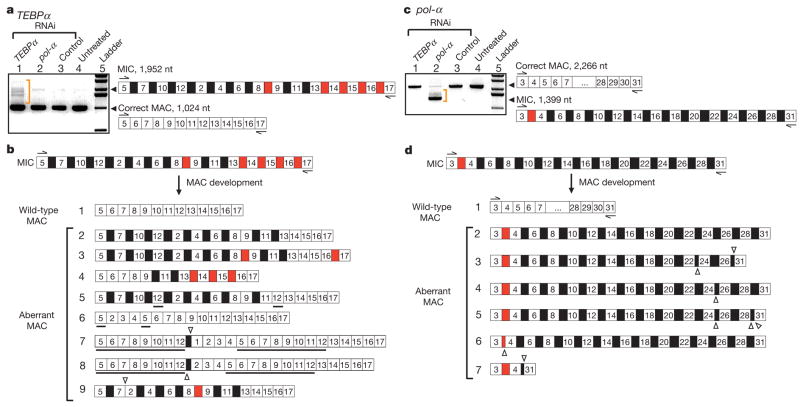
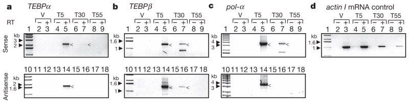
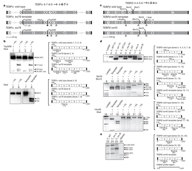
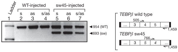
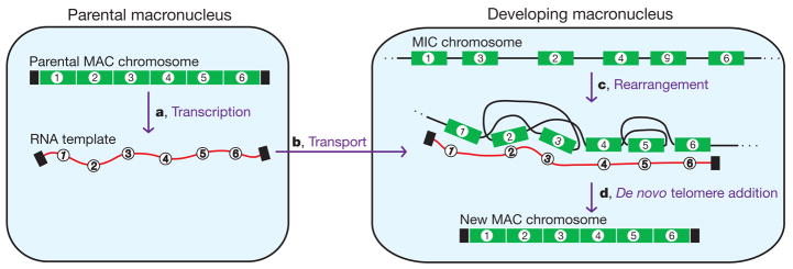
Genome-wide DNA rearrangements occur in many eukaryotes but are most exaggerated in ciliates, making them ideal model systems for epigenetic phenomena. During development of the somatic macronucleus, Oxytricha trifallax destroys 95% of its germ line, severely fragmenting its chromosomes, and then unscrambles hundreds of thousands of remaining fragments by permutation or inversion. Here we demonstrate that DNA or RNA templates can orchestrate these genome rearrangements in Oxytricha, supporting an epigenetic model for sequence-dependent comparison between germline and somatic genomes. A complete RNA cache of the maternal somatic genome may be available at a specific stage during development to provide a template for correct and precise DNA rearrangement. We show the existence of maternal RNA templates that could guide DNA assembly, and that disruption of specific RNA molecules disables rearrangement of the corresponding gene. Injection of artificial templates reprogrammes the DNA rearrangement pathway, suggesting that RNA molecules guide genome rearrangement.


Figures

Figure 1
Figure 1. RNAi against putative RNA templates leads to disruption of DNA rearrangement, with accumulation of aberrant products
MDS segments (white boxes) and IES regions (black boxes if located between nonconsecutive segments, red if consecutive) are drawn schematically, not to scale. The ladders are 1 kb (Invitrogen). a, PCR amplification of the TEBPα region between segments 5 and 17 from total DNA extracted from the sources treated with: TEBPα RNAi, pol-α RNAi and control double-stranded (ds)RNA (184 nucleotide (nt) dsRNA from feeding vector polylinker), as well as untreated cells. Only cells treated with TEBPα RNAi contain partial or incorrect rearrangements, on the basis of size (orange bracket). Cells treated with TEBPα RNAi were fed with dsRNA covering the region between segments 1 and 16. MAC, macronucleus; MIC, micronucleus. b, The sequence of several TEBPα PCR products between segments 5 and 17 in cells treated with TEBPα RNAi. IESs between both scrambled (black) and nonscrambled (red) MDSs are deleted from some molecules at both correct and incorrect (cryptic) repeats. Open triangles show the locations of cryptic junctions between neighbouring segments (if pointing up) and non-neighbouring segments (pointing down) on the basis of the precursor micronuclear order. Underlined segments are duplications. c, PCR amplification of pol-α between segments 3 and 31 from total DNA extracted from sources treated with: TEBPα RNAi, pol-α RNAi and control dsRNA (as above), as well as untreated cells. Only cells treated with pol-α RNAi show aberrantly rearranged products, on the basis of size (orange bracket). Cells treated with pol-α RNAi were fed with dsRNA covering the region between segments 16 and 29. d, Sequence of several pol-α PCR products between segments 3 and 31 in cells treated with pol-α RNAi.
Figure 2
Figure 2. Long sense and antisense transcripts are present during early development
RT–PCR of both strands from cells in a vegetative state (V) as well as 5 (T5), 30 (T30) and 55 (T55) hours post conjugation. Filled arrowheads indicate size of relevant markers (1 kb ladder, Invitrogen). Long, intron-containing maternal transcripts appear 5 h post conjugation and disappear 50 h later. The peak of polytene chromosome formation and DNA rearrangements is estimated to occur in this time window. Arrowheads indicate specific products. All non-specific amplification products were confirmed by sequencing to be unrelated. a, b, c, RT–PCR detection of both sense (+) and antisense (−) strands of TEBPα, TEBPβ and pol-α RNA templates, respectively. d, RT–PCR of actin I mRNA as a control for RNA in each sample.
Figure 3
Figure 3. Microinjection of alternative DNA templates produces alternatively rearranged chromosomes
a, Top, wild-type TEBPα macronuclear chromosome (labelled TEBPα wild type) with segments 1–17 colinear; middle, microinjected TEBPα template designed to switch (sw) the order of segments 7 and 8 (TEBPα sw78 template); bottom, map of resulting macronuclear product (TEBPα sw78), to scale. Asterisks, point mutations in synthetic templates; black rectangles, telomeres; open triangles, cryptic pointers to switch segment order; filled triangle, an unspliced 5 bp IES (see Supplementary Fig. 1). b, Left, PCR and restriction analysis of DNA microinjection products (ethidium bromide staining). Inj., injected. Right, single-sided arrows, PCR primers (in bp); scissors, restriction sites (in bp). Lanes 1–4 show presence of wild-type (WT) macronuclear product as well as a product from microinjected cells with segments 7 and 8 switched (TEBPα sw78; lanes 2, 4). Lanes 5–8 distinguish the microinjected template (lanes 7, 8) from the macronuclear product (lanes 5, 6). c, Top, wild-type TEBPβ macronuclear chromosome (TEBPβ wild type) with segments 1–7 colinear; middle, microinjected template designed to switch the order of segments 4 and 5 (TEBPβ sw45 template); and bottom, map of expected macronuclear product (TEBPβ sw45). d, PCR and restriction analysis of DNA microinjection products. Lanes 1–8 use a simple PCR length assay for the presence of a smaller product when the order of segments 4 and 5 has been reversed (lower band): one week (F1 sw-1w) and two weeks (F1 sw-2w) after microinjection, as well as the putative F2 generation (epigenetic inheritance was also observed for putative F3, see Supplementary Fig. 2; 1 kb DNA ladder (Invitrogen)). F2 sw-A and F2 sw-B are the asexual progeny of two independent conjugating pairs in the F1; sw indicates injected cells or their progeny and WT indicates wild-type non-injected controls. Lanes 9–15 confirm the presence of the macronuclear product in which segments 4 and 5 are switched (TEBPβ sw45) in F1 (one week post injection). Lanes 16–22 distinguish the microinjected template from the F1 (one week) macronuclear product; arrowheads in lanes 19 and 20 point to aberrantly rearranged molecules lacking segment 5. e, Lanes 23–26, HindIII and BsrGl Southern analysis of total DNA extracted from the putative F2 generation.
Figure 4
Figure 4. Microinjection of alternative RNA templates leads to alternatively rearranged chromosomes
RNA microinjection of TEBPβ sw45 template. Sense (s), antisense (as), and combined sense and antisense (s/as) RNA templates were microinjected in both wild-type (control) and switched orientations. Lanes 5–7 display the expected macronuclear product if segments 4 and 5 have been switched (lower band). Primers are as in Fig. 3d. Lane 1, 1 kb ladder (Invitrogen).
Figure 5
Figure 5. Model for RNA guiding of genome rearrangements during macronuclear development in Oxytricha.
a, Bidirectional RNA transcription of all DNA nanochromosomes (including injected DNA) in the old, maternal macronucleus (MAC) before its degradation. b, Transport of these RNA transcripts to the newly developing macronucleus, where they may act as scaffolds to guide rearrangements (deletion, permutation and inversion) of corresponding micronuclear (MIC) DNA sequences (c). This step would be notable and unprecedented, but perhaps possible if there were either local or extensive strand-separation of both the RNA template and the developing DNA (see ref. 20). In this illustration, segments 2 and 3 are switched and segment 5 is inverted (number upside down). d, De novo telomere addition (black rectangles) and amplification completes formation of new macronuclear nanochromosomes.

Comment in

  • Molecular biology. RNA rules.
    Yao MC. Yao MC. Nature. 2008 Jan 10;451(7175):131-2. doi: 10.1038/451131a. Nature. 2008. PMID: 18185572 No abstract available.

References

    1. King ML, Messitt TJ, Mowry KL. Putting RNAs in the right place at the right time: RNA localization in the frog oocyte. Biol Cell. 2005;97:19–33. - PubMed
    1. Tadros W, Lipshitz HD. Setting the stage for development: mRNA translation and stability during oocyte maturation and egg activation in Drosophila. Dev Dyn. 2005;232:593–608. - PubMed
    1. Tang F, et al. Maternal microRNAs are essential for mouse zygotic development. Genes Dev. 2007;21:644–648. - PMC - PubMed
    1. Rassoulzadegan M, et al. RNA-mediated non-mendelian inheritance of an epigenetic change in the mouse. Nature. 2006;441:469–474. - PubMed
    1. Herbert A, Rich A. RNA processing and the evolution of eukaryotes. Nature Genet. 1999;21:265–269. - PubMed

Publication types

MeSH terms

Associated data